The Internal Pterygoid Branch
Description
This section is from the book "Anatomy Of The Arteries Of The Human Body", by John Hatch Power. Also available from Amazon: Anatomy of the Arteries of the Human Body, with the Descriptive Anatomy of the Heart.
The Internal Pterygoid Branch
On reaching the anterior margin of the internal pterygoid muscle the facial artery gives off a small branch which is distributed to the substance of this muscle.
The artery in its facial stage usually gives off the six branches already enumerated: these may be divided into external, internal, and terminating. The buccal and some small muscular branches constitute the external; the inferior labial, the two coronaries, and the dorsalis nasi compose the internal, and the angular is the terminating artery.
The Buccal Branch
The Buccal Branch runs backwards from the outer part of the facial over the buccinator muscle, and then, getting on the inside of the ramus of the lower jaw, terminates by anastomosing with the internal maxillary. Its branches are distributed to the buccinator and masseter muscles, to the fat of the cheek, the parotid gland, and Steno's duct.
The Inferior Labial Branch
The Inferior Labial Branch is distributed to the muscles and integuments of the lower lip, and anastomoses with the submental and dental arteries.
The Inferior Coronary Artery
The Inferior Coronary Artery passes inwards in a very tortuous manner beneath the triangularis oris, and quad-ratus menti, and proceeds along the margin of the lower lip, close to its mucous membrane, where it anastomoses with the artery of the opposite side. In its course it supplies the above-mentioned muscles, and anastomoses with the inferior labial, submental, and dental arteries.
The Superior Coronary Artery
The Superior Coronary Artery arises near the commissure of the lips, and runs tortuously inwards between the labial glands and mucous membrane of the upper lip. On the middle line it anastomoses with the artery of the opposite side, and sends upwards, towards the septum of the nose, a small branch termed the artery of the filtrum, the branches of which are distributed to the muscles, integuments, and mucous membrane of the upper lip and to the gums, where these small vessels anastomose with branches of the inferior dental artery.

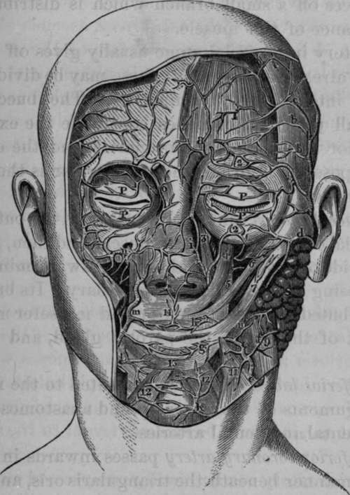
Fig. 13. Dissection of the anastomosis between the Facial, Transverse Facial, branches of the Internal Maxillary, Ophthalmic, and Temporal Arteries.
1, Frontal portion of Occipito-frontalis Muscle. 2, 2. Orbicularis Palpebrarum. 3, 4, Levator Labii Superioris Alaeque Nasi. 5, Levator Anguli Oris. 6, Zygomaticus Minor. 7, Zygomaticus Major. 8. Parotid Gland. 9. Masseter. 10, Small Artery to Buccinator Muscle. 11, Depressor Anguli Oris. 12, 12, Quadratus Menti of each side. 13, Orbicularis Oris. 14, Artery of the Filtrum coming off from the junction of the Superior Coronaries. K, Ascending branch of Submental Artery. P, P, P, P, Palpebral, a, Frontal Artery, b, b, c, c, Branches of Temporal Artery, the upper branch anastomosing with a branch of the Frontal Artery, d, Transversalis Faciei Artery, e, e, Facial or External Maxillary Artery, f. Twig to Masseter Muscle, g, Inferior Coronary Artery, h, Superior Coronary Artery. i, Anastomosis between the Nasal branch of the Ophthalmic and Angular Arteries. 1, Inferior Labial Arterv. m, Facial Artery giving off Superior Coronary Artery, n, Infraorbital Artery, o, Portion of Corrugator Supercilii Muscle.
Continue to:
- prev: The Inferior Mental, Or Submental Branch
- Table of Contents
- next: The Dorsalis Or Lateralis Nasi Artery
