The Facial Artery
Description
This section is from the book "Anatomy Of The Arteries Of The Human Body", by John Hatch Power. Also available from Amazon: Anatomy of the Arteries of the Human Body, with the Descriptive Anatomy of the Heart.
The Facial Artery
The Facial Artery, called also the labial or external maxillary, arises immediately above the lingual, and often together with it by a common trunk. The artery may be divided into two stages,a cervical and a facial stage; in its cervical stage it ascends, lying near the outer surface of the mylo-hyoid and hyo-glossus muscles and under cover of the skin and superficial fascia, platysma myoides, cervical fascia, digastric and stylo-hyoid muscles, the lingual nerve, and portion of the submaxillary gland, into the substance of which it penetrates : in this situation it lies under cover also of the body of the inferior maxillary bone, and, after passing through the gland, touches the internal pterygoid muscle: it here makes a turn, the convexity of which is directed upwards and lies anterior and external to the tonsil: from this point it descends, reaches the inferior margin of the body of the bone and curves underneath its cutaneous surface where the first stage terminates. In its facial stage it ascends tortuously from the inferior margin of the body of the inferior maxilla, along the side of the face, till it arrives at the internal angle of the eye, where it terminates in anastomosing with the nasal and frontal branches of the ophthalmic artery. In this stage it lies on the inferior maxillary bone, in a groove frequently provided for its reception, between the masseter muscle posteriorly, and the triangularis oris anteriorly: it next lies on the buccinator muscle, the levator anguli oris or musculus caninus, the levator labii superioris proprius; and, lastly, on the nasal division of the levator labii superioris alaeque nasi. In this stage it is covered by the skin and superficial fascia, platysma, and frequently by a few of the posterior fibres of the triangularis oris muscle; by the zygomaticus major and minor, by the labial division of the levator labii superioris aloaque nasi near its insertion, and finally by a few of the internal and inferior fibres of the orbicularis palpebrarum muscle. In this situation the artery may be seen, after it has escaped from under cover of the labial portion of the levator labii superioris alaeque nasi, lying against the outer side of the nasal portion of this muscle and thus separating the two portions from each other.
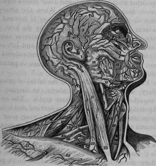
Fig. 12. Dissection of some of the terminating branches of the External Carotid Artery and part of the course of the Subclavian Artery.
A, Right Subclavian Artery in third stage. B, Internal Carotid Artery. C, External Carotid Artery. K, Temporal Artery dividing lower down than usual, a, Supra-scapular Artery crossing Anterior Scalenus Muscle, b, Irregular Posterior Scapular Artery coming from Subclavian, and in this case passing between branches of Brachial Plexus, c, Muscular Artery, e, Superior Thyroid Artery, f, Facial Artery, g, Branch of Transverse Artery of face, h, Branch of Posterior Auris Artery, i, Branch of Occipital Artery. l. Anterior branch of Temporal Artery, m, Posterior branch of Temporal Artery, n, Frontal Artery. l, 1, Pinna. 2, 2, Temporal Muscle covered by Temporal Aponeurosis. 3, Orbicularis Palpebrarum. 4. Angular Artery. 5, Levator Labii Superioris. 6, Levator Anguli Oris, or Musculus Caninus. 7, Zygomaticus Minor. 8, Zygomaticus Major. 9, Orbicularis Oris. 10, Muscular branches of Mental Artery. 11, Depressor Anguli Oris, or Triangularis Oris. 12, Buccinator Muscle. 13, Parotid Gland. 14, Masseter Muscle. 15, Sterno-mastoid Muscle. 16 Muscular branch of Occipital Artery. 17, Submaxillary Gland. 18, Levator Anguli Scapulae Muscle. 19. Middle and Posterior Scaleni Muscles. 20, Anterior belly of Omo-hyoid Muscle. 21. Sterno-thy-roid Muscle. 22, Sterno-hyoid Muscle. 23, Thyroid Cartilage. 24, Trapezius Muscle. 25, Posterior belly of Omo-hyoid Muscle. 26, 26, 26, Brachial Plexus. 27, Anterior Scalenus Muscle. 28, 29, Origins of Sterno-mastoid Muscle. 30, Trachea. 31, Deltoid. 32, Pectoralis Major.
Continue to: