The Internal Carotid Artery
Description
This section is from the book "Anatomy Of The Arteries Of The Human Body", by John Hatch Power. Also available from Amazon: Anatomy of the Arteries of the Human Body, with the Descriptive Anatomy of the Heart.
The Internal Carotid Artery
This artery may be exposed in the following manner: The brain should first be removed in the usual way, leaving uninjured, however, the cerebellum, medulla oblongata, and pons Varolii: the tentorium should now be removed, and the cerebellum pushed gently forward, or a small portion of its posterior part removed, so as to make room for the saw. A vertical section of the cranium should be next made through the posterior part of the occipital foramen and through the cervical vertebrae, behind their articular processes. This section will enable the student to study the medulla oblongata, vertebral arteries and their branches, and the eighth, ninth, and sub-occipital nerves. After these parts have been examined, the cerebellum and spinal marrow may be removed, and the ligaments divided which connect the occipital bone to the first and second vertebrae. The vertebrae may now be separated from the occipital bone, the recti capitis antici muscles having been previously detached from the front of the spine, but allowed to remain in connection with the occipital bone. Lastly, the lower part of the neck may be cut across, and the digastric and styloid muscles, etc. neatly dissected. The tube may be seen if a metallic cast of the ear be previously taken, and the bone softened in dilute acid.
* Med. Chirurg. Trans., vol. viii. 11* portion of the internal carotid contained in the osseous canal must be carefully followed with a chisel, and its exact relation to the cochlea, tympanum, and Eustachian.

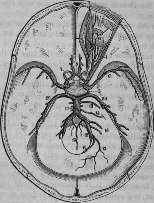
Fig. 16. Arteries of the Interior of the Cranium.
1, Internal Carotid Arteries. 2, Ophthalmic Artery. 3, Posterior communicating Arteries. 4, Anterior Cerebral Arteries. 5, Auterior communicating; Artery. 6, Middle Cerebral Arteries. 7, Lachrymal. 8, Short Ciliary Arteries piercing the back part of the Eyeball. 9, Central Retinal piercing the Optic Nerve to reach the interior of the Eyeball. 10, Muscular Artery. 11, Frontal and Nasal Artery. 12, Vertebral Arteries. 13, Posterior Meningeal Artery. 14, Posterior Spinal Artery. 15, Anterior Spinal Arteries conjoining in a single one. 16, Inferior Cerebellar Arteries. 17, Basilar Artery formed by the union of the Vertebrals. 18, Internal Auditory. 19, Superior Cerebellar. 20, Posterior Cerebral Arteries.
Continue to:
