The Palpebral Arteries
Description
This section is from the book "Anatomy Of The Arteries Of The Human Body", by John Hatch Power. Also available from Amazon: Anatomy of the Arteries of the Human Body, with the Descriptive Anatomy of the Heart.
The Palpebral Arteries
The Palpebral Arteries are two in number; they arise at the inner surface of the optic nerve: the inferior descends behind the tendo oculi, and after sending some twigs to the lachrymal sac, divides into two branches, one of which supplies the inferior division of the orbicularis palpebrarum, while the other follows the adherent margin of the lower tarsal cartilage, and supplies this cartilage, the Meibomian glands, the conjunctiva and skin. The superior palpebral artery arises a little more in front, and after supplying the caruncula lachrymalis, is distributed in the upper eyelid, exactly as those of the inferior artery are in the lower: it anastomoses externally with the lachrymal and temporal arteries.
The terminating branches of the ophthalmic artery are the frontal and the nasal.
The Frontal Artery
The Frontal Artery, usually smaller than the nasal, advances to the superior and internal part of the base of the orbit, from which it escapes in passing between the tendo oculi and pulley of the superior oblique muscle. It then ascends on the forehead, between the frontal bone and orbicularis palpebrarum, and subdivides, to supply this muscle, and the occipito-frontalis and corru-gator supercilii.
The Nasal Artery
The Nasal Artery is larger than the preceding, and with it escapes from the orbit between the tendo oculi and pulley of the superior oblique muscle: it then descends on the side of the root of the nose, and supplies the lachrymal sac, and adjacent muscles, and anastomoses with the termination of the labial or facial artery. In many cases the nasal artery seems to be perfectly continuous with the angular branch of the facial.
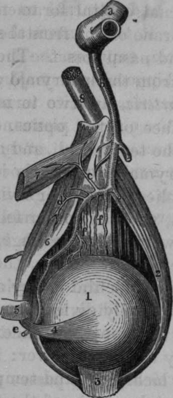
Fig. 18. Branches of Ophthalmic Artery given off under the Superior Rectus Muscle.
1. Ball of the Eye. 2, External Rectus Muscle. 3, Insertion of Superior Rectus Muscle, cut and turned forwards. 4, Tendon of Superior Oblique Muscle which passes underneath the Superior Rectus. 5, Trochlea or Pulley for Superior Oblique Muscle. 6, Belly of Superior Oblique Muscle. 7, Superior Rectus Muscle divided. 8. Optic Nerve, a, Turn of Internal Carotid Artery giving off the Ophthalmic Artery, b, Ophthalmic Artery, c, A twig to Superior Rectus Muscle, d, Muscular Branches, e, Continuation of Ophthalmic Artery cut across, f, f, Some of the short Ciliary Arteries.
In the operations for extracting the eye, the trunk of the ophthalmic is divided, and its sheath prevents it from retracting so as to bleed into the cavity of the cranium; the hemorrhage into the cavity of the orbit is however, frequently very considerable.
After the ophthalmic, the next branch given off by the internal carotid is the choroid artery.
The Choroid Artery
The Choroid Artery is a small but constant branch. It arises from the posterior part of the internal carotid, and passes backwards and outwards towards the crus cerebri: in its course it lies internal to and under cover of the internal convolution of the base of the middle lobe of the brain, and external to the posterior communicating artery: it then enters the inferior cornu of the lateral ventricle, supplies the tractus opticus and crus cerebri, the hippocampus major, pes hippocampi, and corpus fimbriatum, and its terminating branches are distributed to the choroid plexus.
Continue to: