The External Circumflex Artery
Description
This section is from the book "Anatomy Of The Arteries Of The Human Body", by John Hatch Power. Also available from Amazon: Anatomy of the Arteries of the Human Body, with the Descriptive Anatomy of the Heart.
The External Circumflex Artery
The External Circumflex Artery arises from the external side of the profunda, where the latter is forming its curvature in order to descend inwards: from this origin it runs almost transversely outwards behind the sartorius and rectus muscles, and through the midst of the fasciculus of branches descending from the anterior crural nerve. It terminates in three branches: an ascending, transverse, and descending. The ascending branch, considerably the smallest, runs upwards and outwards behind the tensor vaginae femoris, in the interval between the iliacus internus and glutaeus medius muscles, till it reaches the anterior superior spine of the ilium, where it terminates in anastomosing with the superficial and deep circumflexa ilii arteries, and with the glutaeal and ilio-lumbar. The transverse branch, larger than the preceding, runs outwards, in front of the superior extremity of the shaft of the femur, and then curves round to its posterior surface: in this course it passes through the superior fibres of the vastus externus, and then pierces the insertion of the glutaeus maximus. On raising the latter muscle, the termination of this branch is seen: it supplies the adductor muscles, the vastus externus, and the capsule of the hip-joint. The descending branch (or rather set of branches, as there are usually two and frequently more) is much the largest; it runs downwards and outwards, first between the rectus muscle and cruraeus, and then between the tastus externus and cruraeus: it sends many branches to these muscles, and terminates near the patella in inosculation with the anastomotic and external articular arteries. When there is but one descending branch, it goes to the vastus externus. This branch is sometimes greatly enlarged in cases of popliteal aneurism; and in amputations of the thigh it frequently requires the application of a ligature.
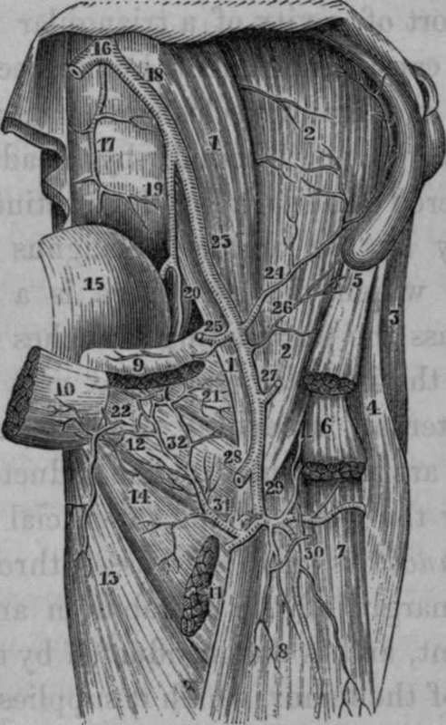
Fig. 61. Represents the Anastomosis between the Obturator and Internal Circumflex Arteries.
1, 1, Psoas Magnus Muscle. 2, 2, Iliacus Internus Muscle. 3. Glutaeus Medius Muscle. 4, Tensor Vaginae Femoris Muscle. 5, Origin of Sartorius Muscle. 6, Portion of Rectus Femoris Muscle. 7, Vastus Externus. 8, Cruneus. 9. Origin of the Pectineus Muscle. 10, Origin of Adductor Longus. 11, Insertion of preceding Muscle into the middle third of the Linea Aspera. 12. Obturator Externus Muscle. 13, Adductor Magnus Muscle. 14, Adductor Brevis Muscle. 15, Urinary Bladder. 10, Division of Abdominal Aorta. 17. Middle Sacral Artery. 18. Left Common Iliac Artery. 19, Internal Iliac Artery. 20. Obturator Artery. 21, Capsular Ligament of hip joint. 22, Muscular twig to the Adductors. 23, External Iliac Artery. 24, The Internal Circumflexa Ilii Artery. 25, Epigastric Artery, cut. 26, External Circumflexa Ilii Artery. 27, Superficial Epigastric Artery, cut. 28, Femoral Artery, cut. 29, Profunda Artery. 30, Descending branches of External Circumflex Artery. 31, Internal Circumflex Artery. 32, Anastomosis between Internal Circumflex and Obturator Arteries.
Continue to: