The Obturator Artery
Description
This section is from the book "Anatomy Of The Arteries Of The Human Body", by John Hatch Power. Also available from Amazon: Anatomy of the Arteries of the Human Body, with the Descriptive Anatomy of the Heart.
The Obturator Artery
This is the smallest and most anterior of the four branches of the internal iliac which go out of the pelvis, and should be dissected before the pudic.
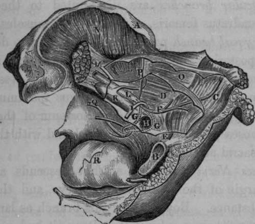
Fig. 46. Represents the Surgical Anatomy of the Obturator Artery, in both its Normal and Abnormal Course, in connection with Femoral Hernia.
A, Anterior Superior Spine of the Ilium. B. Symphysis Pubis. 0, The Rectus Muscle. D, Tho Peritoneum. E, Conjoined Tendons. V, Epigastric Artery. G. G, Two different courses of the Obturator Artery, when given off by the Epigastric. H, Crural Ring. I, Round Ligament of the Uterus. K, External Iliac Vein. L. External Iliac Artery. M, Tendon of Psoas Parvus Muscle, resting on Psoas Magnus. N. Ulacus luternus Muscle. O, Transversalis fascia. P. Circumflexa Ilii Artery. Q. Normal course of Obturator Artery. R, The Urinary Bladder. (See Varieties of the Obturator artery).
It runs downwards and forwards below and within the brim of the true pelvis, in order to pass through the upper part of the obturator foramen. In this course it is accompanied by the obturator nerve which lies above, and the obturator vein which lies beneath it: it communicates with the artery of the opposite side by a branch crossing transversely behind the body of the pubis. When the obturator artery arises from the epigastric, its vein and nerve lie below it. Having passed through the obturator canal, it lies on the obturator externus muscle, covered by the pectineus, and there divides into two branches, an anterior and posterior.
Continue to: