98. The Main Air Tube, The Trachea Or Windpipe
Description
This section is from the book "Animal Physiology: The Structure And Functions Of The Human Body", by John Cleland. Also available from Amazon: Animal Physiology, the Structure and Functions of the Human Body.
98. The Main Air Tube, The Trachea Or Windpipe
The Main Air Tube, The Trachea Or Windpipe, is about four and a half inches long, and is surmounted by the larynx, the part in which the glottis is placed, and which constitutes the organ of voice. The constant patency of the trachea is insured by a series of cartilaginous hoops, which in some animals form complete rings, but in the human subject are deficient behind. This tube divides below into a right and left bronchus of similar structure; and each of these divides and subdivides into smaller and smaller bronchial tubes, the larger of which have crescentic cartilages arranged in their walls, not, as in the trachea, one directly over another, but in such a way as to maintain the cylindrical form of the tube on every aspect, while in the smaller these cartilages degenerate to irregular nodules, and disappear.
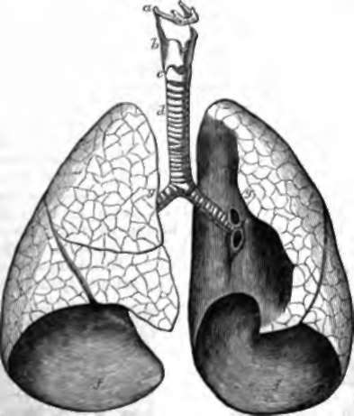
Fig. 74. Human Windpipe and Lungs. a, Hyoid bone; b, c, thyroid and cricoid cartilages of larynx; d, trachea dividing interiorly into right and left bronchus; e, root of left lung, the pulmonary artery and vein cut across; f, f, the bases of the lungs, which rest on the diaphragm; g, g, portions of the anterior margins, which in the booty reach to the middle line, and have only folds of pleura between them. The right lung is seen to have three lobes, the left two; the right is shorter than the left, and the anterior part of the left is hollowed out opposite the position of the apex of the heart.
In all these tubes, except the smallest, there is a longitudinal arrangement of elastic fibrous tissue, enabling them to elongate when required by the stretching of the neck or the expansion of the lungs; also transverse muscular fibres, confined, in the trachea and bronchi, to the back part where the cartilages are deficient, but circular in the other tubes, and capable by their contraction of modifying the freedom of entrance of air into the lungs, as is strikingly exemplified in asthma, which consists of spasms of these muscles to such extent as to produce difficulty of breathing. The mucous membrane is furnished with mucous glands, and lined with ciliated columnar epithelium. But the bronchial tubes of smallest size, reduced to inch in diameter, have homogeneous walls with a simple squamous lining, and each of these terminates in an ultimate lobule or infundibulum, consisting, as has been already said, of an irregular passage, surrounded with air cells. These air cells or locules are cup-shaped depressions, consisting of a framework of fine elastic tissue, and one of the closest capillary networks in the body, in which circulates the blood sent to the lungs by the pulmonary artery; and in order to secure the greatest possible contact of the blood with the air, the septa between the locules present only one layer of capillaries exposed on both sides, and protected only by a very delicate epithelium.
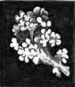
Fig. 75. Cast of Infundibula. 15/1; from Henle.
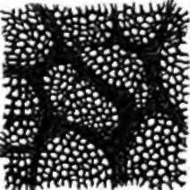
Fig. 76. Air Cells, with capillary blood-vessels injected, 50/1.
The ultimate lobules are united in groups to form larger lobules, from a quarter to half an inch in diameter, very closely united one to another, but with outlines which, in those of the surface of the lung, can be easily distinguished, particularly in the human subject, where they are usually more or less marked by the deposit of black pigment. The tissue between the lobules and that of the bronchial tubes require, like all other tissue, oxygenated blood for their nourishment; and as that which arrives by the pulmonary artery is unfit for use, they receive their supply from small systemic branches called the bronchial arteries.
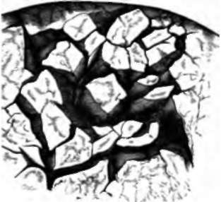
Fig. 77. Lobules of Human Lung. partially separated; natural size.
Each lung is completely invested with a serous membrane, the pleura, the visceral layer of which is adherent to its surface, while the parietal layer is reflected from what is called the root of the lung, where the bronchus and blood-vessels enter, to the walls of the chest, the outer surface of the pericardium, and the upper surface of the diaphragm.
99. The means by which the air is introduced into the lungs and expelled therefrom, is similar to that by which it is drawn into and forced out from a concertina. The interior of the lungs communicates freely with the air outside by means of the windpipe, and when the capacity of the cavity containing them is enlarged, the air passes in, while, when the capacity is diminished, a quantity of air is expelled. This is the principle of the mechanism of respiration in the majority of animals; but in frogs the want of osseous thoracic walls, and in turtles their rigidity, renders it impracticable; and therefore both these animals pump the air down into their lungs by a motion of a swallowing description, which may be seen constantly going on in their throats.
Continue to:
- prev: 97. The Object Of Respiration
- Table of Contents
- next: 100. The Expansion And Diminution Of The Chest In Respiration