90. The Arteries
Description
This section is from the book "Animal Physiology: The Structure And Functions Of The Human Body", by John Cleland. Also available from Amazon: Animal Physiology, the Structure and Functions of the Human Body.
90. The Arteries
The Arteries, into which the blood is sent by the heart, are a series of branching, elastic, and contractile tubes. They have a smooth internal lining, and externally have a tough felted coat of areolar tissue; but the main thickness of their walls consists of a middle coat of elastic and muscular fibres intermixed and arranged circularly, lying among meshes of elastic membrane. In the larger arteries the muscular fibres are exceedingly small, and the elastic fibres abundant; but as the vessels get smaller there is a greater development of muscular tissue, and less of the elastic, until in the minute arterioles the elastic tissue disappears altogether, and the middle coat consists of muscle only.

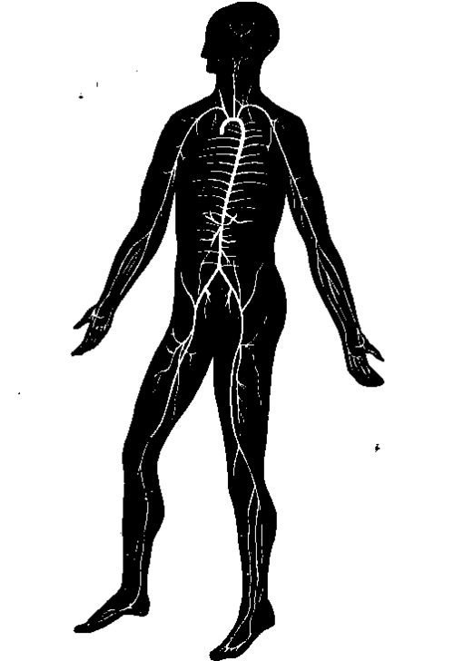
Fig. 67. Arterial System.
The advantage of the elasticity of the arterial walls may be easily illustrated. If a glass tube have a nozzle fastened into it at one end, and at the other be fitted to the stop-cock of a water pipe, and if the water be turned on and off alternately so as to imitate the repeated discharges of blood from the ventricles, the water will emerge from the nozzle in jets, which will cease instantaneously each time that it is turned off. But if the same experiment be made with a long india-rubber tube instead of a glass one, the water will spring from the nozzle in a continuous flow, notwithstanding the interrupted manner in which it is admitted to the tube; and if the experiment be varied so that the glass and the india-rubber tube shall both be filled from the top at the same time, while the nozzles on the two tubes are of the same size, the elastic one will discharge in a given time a much larger quantity of water than the one which is rigid. In the rigid tube there is great loss of force by friction; while in the elastic tube, as each fresh jet of fluid enters, the walls are distended, and as it ceases they recover, and give their contents a fresh propulsion onwards in a second wave, which distends the tube further on; and thus, after traversing a sufficient length of tube, the interrupted stream is converted into one which is continuous. This is precisely what happens in the arteries. When a large artery is divided, the blood comes in separate abrupt jets with well marked intervals between ; in smaller arteries the duration of the jets is longer and the intervals are shorter, and from little twigs the blood spouts out in an almost continuous stream.
While the elasticity of the arteries thus converts the separate gushes of blood from the heart into one continuous flow before the capillaries are reached, their contractility, derived from their muscular fibres, determines the amount of blood which is sent at different times to each part. Their contraction is not of the vermicular description, but purely tonic: they do not assist the forward movement of the blood by propelling it onwards, but, by varying in diameter at different times, they allow more or less blood to pass through them. The muscular fibres are governed by nerves, termed vasomotor: these are found in the sympathetic trunks (p. 215), and their action may be illustrated by dividing the sympathetic nerve of a rabbit in the neck, when immediately the ear of the side experimented on gets red, and the whole of that side of the head becomes warmer than the other; the reason being that the paralysed arterioles no longer resist the entrance of the red and warm blood, but allow it to distend them. This condition is the same as takes place in blushing, only in blushing the withdrawal of the nervous stimulus is temporary, caused by the communication of a disturbing influence from the brain, the result of emotion.
Continue to:
- prev: 89. The Frequency Of The Heart's Pulsations
- Table of Contents
- next: 91. The Pulse In The Arteries
