92. The Capillaries
Description
This section is from the book "Animal Physiology: The Structure And Functions Of The Human Body", by John Cleland. Also available from Amazon: Animal Physiology, the Structure and Functions of the Human Body.
92. The Capillaries
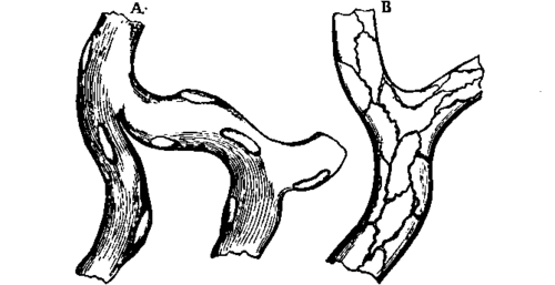
The Capillaries are the smallest blood-vessels, those through the walls of which materials pass to and from the tissues, and so delicate that, as has already been pointed out, even blood corpuscles are able, without injury to the walls, to escape from them into the parts around. They vary from 1/2000 to 1/3500 inch in diameter, and are arranged like the meshes of a net. The meshes vary in size and form in different localities: for the most part they are polygonal; in the papillae of the skin they are in loops; in muscle they are oblong; and in the lung they are circular, with the diameters of the circles little greater than the breadth of the capillaries between which they lie. In some tissues they can be seen under the microscope without previous preparation; and they exhibit the appearance of a homogeneous membranous wall with oval nuclei imbedded in it, and projecting to the outside. With the aid of a weak solution of nitrate of silver, a delicate lining of epithelium, or endothelium as it is sometimes called, is brought into view; but it must not be supposed that the nuclei mentioned belong to that lining.

Fig. 69. Capillaries, highly magnified. A, exhibits the nuclei; B, the endothelium as displayed by means of nitrate of silver.
The blood can be seen circulating in the capillaries of the web of a frog's foot, a tadpole's tail, or a bat's wing, without injury to the animal; and may be still better studied in some internal parts of small animals operated on for the purpose. Furthermore, by gazing steadily at a bright field, and moving a finger rapidly in front of the eye, some persons are able to bring into view the blood corpuscles coursing in the capillaries of the retina in their own eyes (p. 251). From such data as these, the calculation is made that the blood moves in the capillaries in the human subject at the rate of one or two inches per minute. The movement looks much more rapid when seen under the microscope in a frog's foot; but the reason of that is, that the distance which the corpuscles travel being magnified, the apparent rate of motion is proportionately increased; because the rate of movement is the distance travelled in a given time.
Continue to:
