93. The Veins
Description
This section is from the book "Animal Physiology: The Structure And Functions Of The Human Body", by John Cleland. Also available from Amazon: Animal Physiology, the Structure and Functions of the Human Body.
93. The Veins
The Veins begin by radicles from the capillaries, in like manner as the arteries end in these vessels. The blood moves in them from the capillaries towards the heart, and their course on that account is described from twig to trunk like the course of a river. They are larger than the corresponding branches of arteries, and in the limbs they are more numerous. Thus, in the lower limb below the knee, and in the upper limb below the armpit, the main arteries are accompanied with vence comites, that is to say, two or more veins frequently communicating; and there are, besides, large veins beneath the skin without any corresponding artery. The walls of veins are much thinner than arterial walls, owing to their having the middle or muscular and elastic tunic so slightly developed that their principal thickness consists of the external or felted coat; and thus it happens that in a wound, an artery cut across forthwith contracts so as to lessen its apparent diameter, while a vein gapes and continues as large as ever.
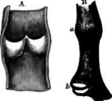
The veins give little or no assistance to the flow of blood by elastic recoil, and, indeed, become easily distended to an undue extent; but they present a peculiar provision to prevent the accumulation of pressure within them, and regurgitation backwards, for they are provided here and there with valves. These valves are on the same principle as the arterial valves of the heart, consisting of semilunar pouches; but the pouches look towards the heart, and instead of there being three of them together, there are only two. They are very delicate, consisting of folds of the lining membrane of the vessel, and are quite transparent; but when liquid is injected from the direction of the heart, it is effectually prevented by them from passing back to the twigs. They are nearly absent from the head and neck, and are most abundant in the lower limbs. By preventing regurgitation, they convert the accidental pressure of surrounding parts into an auxiliary of the circulation, pushing the blood onwards, but incapable of pushing it backwards. Such pressure may, however, easily be sufficiently great to prevent the entrance of blood into a vein; thus in letting blood from the arm it is customary to make the patient exercise the fingers sufficiently to keep up pressure by contraction of the flexor muscles in the forearm on the deep veins, and so compel the return of the blood by the superficial set.

Fig. 70. Venous Valves. A, a vein laid open to show the two pouches of a valve. B, an unopened vein, exhibits at a, the dilatation opposite a valve ; and at b, a closed valve seen from below.
It will be perceived from what has been said, that the rate of flow of the blood in individual veins is very variable. Looking, however, at the venous system as a whole, it will be easily understood that it pours into the heart, in a given time, exactly the same amount of blood as is discharged into the arteries; and as the sectional area of the veins is everywhere greater than that of the corresponding arteries, the flow of blood within them is in the same proportion slower, probably nowhere more than half as fast; but as, in the arteries, the velocity of the blood is less the nearer it approaches the capillaries, on account of the larger sectional area of the total number of vessels in which it is distributed, so it again increases in the passage from the small to the large veins.
Continue to:
- prev: 92. The Capillaries
- Table of Contents
- next: 94. The Pressure Or Force With Which The Blood Is Urged
