11. Elastic Tissue
Description
This section is from the book "Animal Physiology: The Structure And Functions Of The Human Body", by John Cleland. Also available from Amazon: Animal Physiology, the Structure and Functions of the Human Body.
11. Elastic Tissue
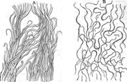
Elastic Tissue occurs both in the form of fibres and thin homogeneous membranes. It gets its name from being highly extensible and resilient, and is most widely distributed in the fibrous form. In the human subject there is only one set of ligaments which consist of nearly pure elastic tissue, namely, the ligamenta subflava, which join together the arches of the vertebra, and get their name from the yellow colour peculiar to elastic fibres. They facilitate, by their resiliency, the resumption of the erect posture when the back has been bent forward. In quadrupeds two other notable instances of pure elastic tissue may be mentioned. One is the ligamentum nuchæ, a strong band extending from the back of the skull, and attaching it to the withers or dorsal spines; the other is in the form of an aponeurosis, which lies on the abdominal wall, and aids the support of the viscera. On examining fibres from any of these sources, it is seen that they may be of considerable breadth, that loosened from their attachments and teased out they curl up at the ends, that they refract light very strongly, and that they are unaltered by acetic acid. Elastic tissue is not easily altered by even prolonged boiling, and yields no gelatin. In many places, as, for example, underneath the pleura, isolated elastic fibres are exceedingly abundant, of great length, curling naturally, and crossing one another in all directions.

Fig. 6. Elastic Tissue. A, From ligamentum nuchæ of sheep. B, From pleural surface of lung.

Fig. 7. Adipose Tissue. A, The usual appearance of fat-cells; a, shows a nucleus on the side of a fat-cell; b, cell filled partly with water, partly with oil. B, Scheme of the mode of accumulation of oil in young fat-cells.
Continue to:
