95. The Time Required For A Portion Of The Blood To Be Carried Through The Whole Circulation
Description
This section is from the book "Animal Physiology: The Structure And Functions Of The Human Body", by John Cleland. Also available from Amazon: Animal Physiology, the Structure and Functions of the Human Body.
95. The Time Required For A Portion Of The Blood To Be Carried Through The Whole Circulation
The Time Required For A Portion Of The Blood To Be Carried Through The Whole Circulation, has been made the subject of most interesting experiments. An easily detected substance, such as ferrocyanide of potassium, is introduced into a vein on one side of the neck of an animal, and the time noted which elapses before it is present in the blood allowed to flow from the corresponding vein on the other side. The substance introduced has to pass through the heart and lungs, and some part of the head or neck, before it can reach the aperture where it is sought for, and thus makes a circuit through both pulmonary and systemic circulation. In the horse, such a circulation is completed in little more than half a minute, and in smaller animals in a much shorter time. Small animals have the pulse rapid; and the rule may be laid down that a complete circulation takes place in from 20 to 30 beats of the heart. This may well appear incredible at first, when it is considered how slowly the blood moves in the capillaries, but the explanation is found by taking into account the exceedingly short distance of capillary circulation traversed by each portion of blood, probably in no case exceeding the tenth of an inch.

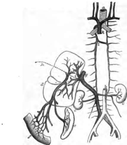
Fig. 71. Venous System, diagrammatic view, a, Trachea dividing into the two bronchi; b, aorta dividing into the two common iliac arteries, which again divide into the external and internal iliac a; c, c, kidneys, with the renal veins emerging from them; d, liver; e, spleen; f, portion of intestine, with mesenteric vein proceeding from it to join the splenic and form the portal vein, which branches in the liver; g, inferior vena cava receiving the hepatic veins emerging from the liver; A, obliterated umbilical vein; i, obliterated ductus venosus; k, superior vena cava formed by union of the two innominate or brachio-cephalic veins; l, the right vena azygos joined by the left.
Continue to:
- prev: 94. The Pressure Or Force With Which The Blood Is Urged
- Table of Contents
- next: 96. Portal System
