The Anterior Tibial Artery
Description
This section is from the book "Anatomy Of The Arteries Of The Human Body", by John Hatch Power. Also available from Amazon: Anatomy of the Arteries of the Human Body, with the Descriptive Anatomy of the Heart.
The Anterior Tibial Artery
This artery is smaller than the posterior tibial: it runs at first somewhat horizontally forwards from the posterior to the anterior region of the leg, through a foramen above the interosseous ligament: this aperture is bounded internally by the tibia, externally by the fibula, which is sometimes grooved by the artery, superiorly by the superior tibiofibular articulation, and inferiorly by the upper fibres of the interosseous ligament, which present a concave margin towards the artery. In this stage of its course the vessel lies close to the fibula, and is occasionally accompanied by a small nerve which connects the posterior with the anterior tibial nerve: it then descends obliquely forwards, and nearly parallel to a line extending from the head of the fibula to the middle line of the ankle-joint. In the rest of its course it has been called by some the Dorsalis Pedis: it runs on the dorsum of the foot to the interval between the metatarsal bones of the great and second toes; here it terminates by dividing into two branches, viz.: the dorsalis pollicis and the arteria communicans.
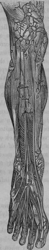
Fig. 65. Represents the Superficial Arteries of the Ante-rior Aspect of the Leg and Foot.
1, The Patella. 2, 3, External and Internal portions of Triceps. 4, Tendon of Rectus. 5, Ligamentum Patellae. 6, External Lateral Ligament of Knee. 7, Bleeps Muscle. 8, Tendon of Sartorius. 9, Tibia. 10, Malleolus Internus. 11, Malleolus Externus. 12, 13, 14, Gastrocnemius and Soleus Muscles. 15, Tibialis Anticus. 16, Long Extensor Muscle of the Toes. 17, Extensor Pollicis Proprius. 18, Peroneus Longus. 19, Peroneus Brevis. 20, Peroneus Tertius or Anticus. 21, 21, 21, Extensor Digitorum Brevis. 22, 22, Inter-ossei. 23, Superior External Articular Artery of Knee. 24, 24, Branch from Superior Internal Articular Artery of Knee. 25, A Superficial Branch from Inferior Internal Articular Artery of Knee. 26, Branch from Inferior External Articular Artery of Knee. 27. 27, Twigs from Anterior Tibial Recurrent. 28, Arterial Anastomosis over the Patella. 29, 29, 29, Superficial branches from Anterior Tibial Artery. 30, Anterior Peroneal Artery. 31, Anterior Tibial Artery. 32, Anterior External Malleolar Artery. 33, Twig from Posterior Internal Malleolar Artery. 34, Twigs from Anterior Internal Articular Artery. 35, Dorsal Artery of Foot. 36, Tarsal Artery. The dotted lines intended to show its course through the fibres of the short Extensor of the Toes. 37, The Dorsalis Pollicis.
In its course down the front of the leg its posterior surface rests, first, on a few fibres of the tibialis posticus which accompany the artery through the opening; then on the interosseous ligament, next on the anterior surface of the inferior extremity of the tibia, and lastly on the astragulus, scaphoid, and internal cuneiform bones: its anterior surface is covered by the anterior tibial nerve, and by the annular ligament; lower down it is crossed by the tendon of the extensor pollicis longus; and near its termination, by the internal tendon of the extensor brevis digitorum: its internal surface corresponds, in the greatest part of its extent, to the tibialis anticus muscle: the external surface is applied, superiorly, to the fibres of the extensor longus digitorum, from which it is separated lower down by the fibres of the extensor pollicis, the internal surface of which muscle guides the anterior tibial nerve over to the outer side of the artery. The tendon of this last muscle crosses in front of the artery on the dorsum of the foot, to get to its inside, and then the vessel is once more related externally to the extensor longus digitorum. In all this course the artery is accompanied by two venae comites, one on either side. The anterior tibial nerve is a branch of the fibular, which winds round the outside of the head of the fibula, passing through the peroneus longus muscle, and meets the outer surface of the artery near the superior extremity of the extensor pollicis muscle. Thus the nerve is at first external to this vessel, then lies on it or in front of it, and inferiorly gets a little to its inner side.
Continue to:
- prev: The Azygos Or Middle Articular Artery
- Table of Contents
- next: Ligature Of The Anterior Tibial Artery