The Foot
Description
This section is from the book "The Anatomy Of The Human Skeleton", by J. Ernest Frazer. Also available from Amazon: The anatomy of the human skeleton.
The Foot
The skeleton of the foot, like that of the hand, consists of a closely articulated number of irregular bones, the tarsus, carrying five long bones, the metatarsus, which in their turn support the phalanges of the free digits (Fig. 138).
The Tarsus
This comprises seven bones, of which one, the astragalus or talus, articulates with the bones of the leg and rests below on the os calcis or calcaneum, which makes the projection of the heel. The astragalus ends in front in a rounded head, which, being directed somewhat inwards as well as forwards, tends to project over the inner side of the front end of the os calcis, and the latter bone has a projection here to support it, hence termed the sustentaculum tali.
These two bones are the largest in the tarsus and form its hinder part : their anterior extremities, nearly on the same level, are joined at the mid-tarsal joint with the front part of the tarsus.
The head of the astragalus articulates with the scaphoid or navicular, and this has the three cuneiform bones against its anterior surface. The cuneiforms are inner, middle and outer, and support the inner three metatarsals.
The anterior end of the calcaneum has the cuboid articulating with it, and this carries on its front aspect the two outer metatarsals.
We see, then, that the astragalus is continuous with the chain of bones on the inner side of the foot, scaphoid, cuneiforms and metatarsals, while the os calcis, cuboid, and outer two metatarsals form a chain of bones that lies outside and rather below the others.
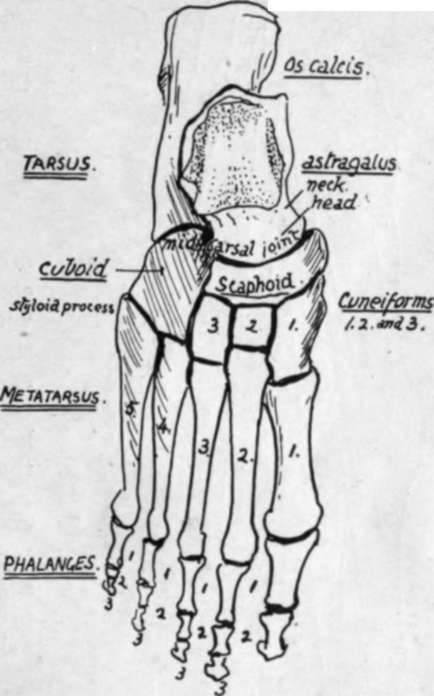
Semidiagrammatic dorsal view of right foot bones.
The Metatarsus
This can be compared with the metacarpus in the hand, when its constituent bones will be seen to be weaker and less divergent. Each bone presents a base, shaft, and head. Notice the obliquity of the line of the tarso-meta-tarsal junction, with the prominent styloid process that projects externally on the base of the fifth metatarsal. The first metatarsal, carrying the big toe, is very thick and strong for supporting the greater part of the weight of the body in stepping off from the foot, whereas the outer portion of the metatarsus is mainly used as a support in balancing the body, and not as a weight carrier, wherefore its comparative weakness.
The Phalanges
As in the hand, these are two in number in the big toe and three in the others, numbered first, second, and third in order from behind forwards in each toe. Notice the thin shafts and thick extremities, save in the great toe, and also observe that there is frequently modification in the direction of fusion or reduction in the last phalanges of the outer toes.
When the names and general relations to each other of the bones of the foot are familiar, the complete skeleton of the region should be studied with a view to understanding the relations and significance of the various bony points, etc., in it, and after that with the object of grasping the means by which the arches of the foot are held in position to support the weight thrown on them.
Looking first at the dorsal surface (Fig. 138), we perceive that the general contour of the tarsus is markedly convex from side to side and also to a slight degree from behind forwards : there are no prominent bony points that make themselves evident above the general level. On this surface, however, there are several things to be observed. We can see that the astragalus consists of a stout body that carries the articular surfaces of the leg bones, and this is joined by a short neck with the rounded head. Under the outer side of the neck is the outer end of a short interosseous canal or tunnel, the sinus tarsi, running obliquely forward and outward between the astragalus and os calcis, and lodging an interosseous hgament connecting the bones : the os calcis has an exposed dorsal surface where this canal opens, and here the anterior annular ligament and Extensor brevis digitorum are attached.
The structures that pass on to the dorsum of the foot from the front of the tibia stream down over the head of the astragalus and mask it somewhat when the living foot is examined from the dorsum. Fig. 139 is a diagram to show the relation between these structures and the underlying bones : they can be made out, in spite of their general smoothness, by careful examination of the living foot.
Looking at the dorsal surface of the anterior tarsal region the shortness of the middle cuneiform is very evident : this gives the bone a square outline, whereas the outer cuneiform has an oblong shape when seen from the top, and this difference enables one to distinguish the two bones at once when they are loose.
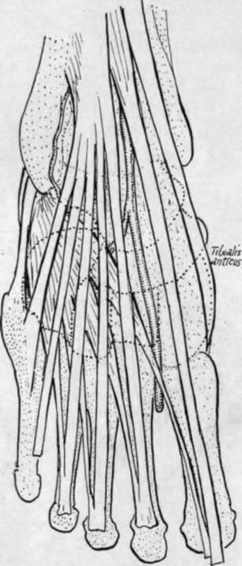
Fig. 139.-To show relation of dorsal structures to the bones.
Notice also how the depth of the cuboid is much less in the outer part, both from above downwards and from before backwards in conformity with the oblique line of the tarsometatarsal joint. Finally, look at the line of this joint more closely and observe that it is irregular, for the inner and outer cuneiforms project forward beyond the line, so that the bases of the first and third metacarpals are in front of the levels of the second and fourth ; or, to call attention to the most noticeable feature, the base of the second metatarsal projects beyond the line of the others in conformity with the small size of the middle cuneiform.
Now examine the lower or plantar surface of the skeleton of the foot. There is a marked concavity on this aspect that is bounded behind by (Fig. 140) the prominent tuberosity of the os calcis : this gives attachment to the superficial muscles of the sole, and the strong plantar fascia, so that an overhanging lip is seen on it, and inner and outer tubercles, the former being the larger. On its inner side, between the tuberosity and the sustentaculum tab, the bone is concave and gives origin to the inner head of Flexor acces-sorius.
Continue to: