Cuneiform Bones
Description
This section is from the book "The Anatomy Of The Human Skeleton", by J. Ernest Frazer. Also available from Amazon: The anatomy of the human skeleton.
Cuneiform Bones
The first or internal cuneiform is the largest, with a heavy thick base below and a thin edge above. Its general appearance distinguishes it at once from the other cuneiforms, of which the second or middle bone can be recognised immediately by the square cut of its dorsal surface or base, which contrasts with the oblong dorsal surface of the third or outer bone (Fig. 138). This figure also shows that the second metatarsal fits in between the inner and outer cuneiforms, so that these bones will have side facets for this metatarsal in addition to the anterior surfaces for their proper metatarsals: also the outer bone comes a little further forward than the cuboid, and consequently has another facet on its outer side for the fourth metatarsal.
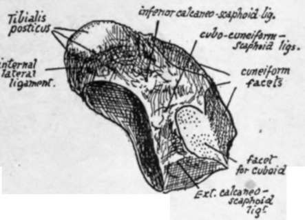
Fig. 150.-Plantar view of left scaphoid. From a foot which exhibited articulation between scaphoid and cuboid.
Inner Cuneiform
A triangular facet behind, with base below, for scaphoid, and a larger kidney-shaped surface in front for the first metatarsal. Inner surface convex from above downwards, conforming with the rounded inner border of the foot, marked by ligaments, covered by skin and superficial tissues, and crossed by tendon of Tibialis anticus, which is partly inserted into it : an oval facet below and in front is partly for its insertion and partly for a bursa under a sesamoid cartilage in the fibres that pass over it to reach the first metatarsal.
The outer surface is on the whole concave. It has an elongated facet along its back and upper margins for the second cuneiform and a small surface for the second metatarsal in its upper and front corner, continuous with the last ; otherwise this aspect is rough for ligamentous fibres (interosseous) of the transverse set joining it to the second cuneiform and metatarsal, but an impression in its lower and front part frequently shows insertion of Peroneus longus. The upper and front ligamentous fibres constitute " Lisfranc's ligament".
The thick base is covered by ligamentous fibres and expansions from the tendon of Tibialis posticus : superficial to these lie the fleshy mass of the short flexor and tendon of long flexor of the big toe and the more superficial structures, and the bone rests through these on the ground when sufficient weight is thrown on the foot to depress the inner arch to a slight extent.
Middle Cuneiform
-Triangular facet in front for second metatarsal and behind for scaphoid. Dorsally it has slight ligamentous markings. Internally a long reshaped facet along the back and upper margins for the inner cuneiform : the rest of the surface for attachment of intercuneiform ligaments of the transverse set. Externally a long facet, slightly narrowed in the middle, along its posterior margin, for the outer cuneiform : the remaining surface ligamentous.
Outer Cuneiform
-Dorsal surface has shght ligamentous markings. Posterior facet for scaphoid does not take up the whole of this aspect, as a blunt point of bone projects slightly below it. Internally a long facet behind for middle cuneiform, and another, usually divided, along the front border for second metatarsal : the remaining surface ligamentous. Externally a rounded facet for cuboid behind and above, a small one for fourth metatarsal * in front and above, and the rest of the surface rough for ligaments of the transverse groups. It carries the third metatarsal alone on its front aspect.
The cuneiforms show a slight general convexity from side to side on their dorsal aspect, where they are covered by the extensor tendons, under which the middle bone is crossed by the dorsal vessels and nerve.. The plantar edges of the two outer bones are covered by ligamentous fibres, from which arise fibres of the Adductor obliquus and some Interosseous muscles.
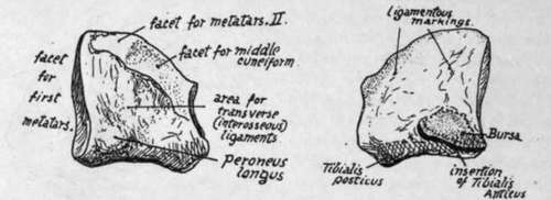
Fig. 151.-Internal cuneiform, left side. Inner and outer views.
* The connection is frequently ligamentous, when no facet is present.
Continue to:
- prev: Scaphoid Or Navicular
- Table of Contents
- next: Metatarsus