The Neck
Description
This section is from the book "The Anatomy Of The Human Skeleton", by J. Ernest Frazer. Also available from Amazon: The anatomy of the human skeleton.
The Neck
The neck is really a prolongation of the shaft, in its development and ossification and in its structure. At birth (Fig. 116) the neck is short and thick and ossified by the extension upwards of the shaft ossification, capped by the cartilaginous head. As development proceeds the neck is gradually elongated, still carrying the epiphysis of the head as a cap on its extremity. The angle it forms with the shaft varies with the extent of its growth, being more open in the young bones and decreasing as growth proceeds, but not after full growth is attained. In the adult the angle is as a rule about 120 degrees, but may vary between no degrees and 140 degrees : it is smaller in women than in men and in short than in long bones. Modification of the angle is largely brought about by the extent of development of the strong ridge on the lower and front part of the neck, that acts as a bracket in strengthening the curve. The neck should be looked on as the upper part of the shaft (Fig. 116) incurved, with the muscular trochanters applied to it, but not interfering with the lines of the walls of the shaft : intersecting arcades of cancellous lamellae spring from these walls and support the articular surface.
The front of the neck is ridged in its long axis and shows numerous vascular foramina. . These occur owing to the fibrous bands with small accompanying vessels that lie under the synovial membrane : the membrane- covers the whole of this face of the bone, extending to the anterior intertrochanteric line. The fibrous bands run from the capsular attachment here towards the head: they are termed retinacula, and there are three main collections of these fibres-below and behind, below and in front, and above, internal to the front part of the great trochanter. The last-mentioned is a broad band that extends in towards the head for some distance beyond the epiphysial line of the trochanter, which (Fig. 117) encroaches here considerably on the upper aspect of the neck.
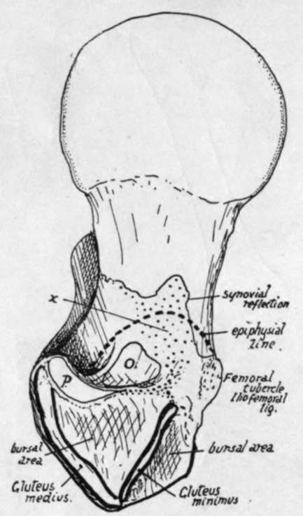
Fig. 117.-Upper aspect of head, neck and great trochanter of right femur. 0. Obturator internus insertion ; P. Pyriformis insertion. The area of insertion of the fibres of the superior retinaculum is marked at x, and it is seen to be continuous with that of Pyriformis round the front of O. and to extend on the neck further than the epiphysial line. Compare with Fig. 118.
The posterior aspect is usually smooth, because there are here no true transverse fibres in the capsule to gain an insertion on the bone : a few marginal fibres of the circular group are the only ones that have any insertion into the femur, and the synovial membrane is reflected from the bone to the deep surface of the circular zone directly. The line of reflection (Fig. 117) is about half-way up the neck, continued up from the turned-up lower end of the anterior intertrochanteric line.
The Obturator externus is closely applied to the lower and back part of the neck, passing to the digital fossa, and the pressure of this muscle moulds the shape of the bone slightly : an indefinite suggestion of an oblique line (Fig. 118) directed outwards and upwards shows the upper limit of the surface affected by this muscle. Below this line the capsule is in relation with the Obturator externus, and above it is covered by the Obturator internus and Gemelli, so that the Quadratus femoris has no direct relation with the capsule at all.
The existence of a long neck is a necessity in a bone which, like the femur, has a head received in a deep articular cavity for purpose of security : if such freedom of movement as the femur enjoys is to be obtained, the body of the bone must be carried at some distance from the embedded head. Necks are more vertical in long femora.
Continue to:
- prev: Head
- Table of Contents
- next: The Great Trochanter