The Anterior Intertrochanteric Line
Description
This section is from the book "The Anatomy Of The Human Skeleton", by J. Ernest Frazer. Also available from Amazon: The anatomy of the human skeleton.
The Anterior Intertrochanteric Line
The anterior intertrochanteric line marks the attachment of the ilio-femoral band : the outer strong limb of the ligament goes to the tubercle at the upper end of the line, the inner band to the lower end of the marking, and the intermediate thinner part to the line in between. The line turns upwards and backwards for a little distance at its lower end, this part of the marking being mostly made by pubo-femoral fibres, and this will indicate the level of the synovial reflection on the back of the neck. The interval between the line and the small trochanter affords insertion to muscular fibres of Iliacus.
* Occasionally also the lowest fibres of Quadratus femoris.
The line appears at first sight continuous with the spiral line made by the aponeurotic origin of Vastus internus on the inner side of the shaft, but closer examination will show that this continuity is only along the outermost part of the intertrochanteric ridge : here the Vastus arises, while the remainder of the ridge is purely ligamentous in origin.
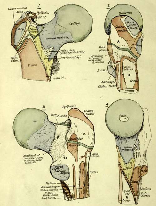
Fig. 118.-Upper end of right femur. The epiphysial line for the great trochanter is marked in green round its base. The retinacula of Weitbrecht," fibres running back toward the head under the synovial membrane, are shown only where they are congregated into their three main groups ; they are derived from the transverse capsular fibres, and the upper one obtains many fibres from Pyriformis (see Fig. 117). I. Anterior aspect. Observe that the Gluteus minimus is attached only to the outer ridge of the trochanter, but its tendon is continuous below with an aponeurotic sheet, the ilio-trochan-teric band, which covers the bursa in front and reaches the bone internal to it. The upper part of the origin of Crureus is mainly tendinous. The extension of the cartilage of the head on to the neck is shown at x ; this lies under the ilio-femoral band or, if the opening for the sub-Psoas bursa is large, under the tendon of the Psoas. 2. From the outer side. The oblique insertion of Gluteus medius is continuous below and in front with that of Gluteus minimus, and frequently with that of Pyriformis above and behind ; it divides this aspect of the trochanter into two areas, one, C, in front and above, under cover of medius and therefore bevelled off in the direction of that muscle, the other, A, below and behind, covered by Gluteus maximus and therefore moulded by that muscle so that it is more vertically directed and curved from before backwards. The surface C carries a bursa, but A has only occasionally an extension of the bursa situated below in relation with it. D, surface covered by Vastus extemus and more or less flattened by it. Crureus fuses with V. externus at a lower level. 3. Posterior aspect. D, surface covered by Quadratus femoris ; deep to this muscle the Obturator externus lies against the bone, moulding the back and lower part of the neck in the area F as it passes to the digital fossa. 4. From the inner side. Observe the pointed area between the spiral line and pectineal line which is occupied by Iliacus. E, inner surface, covered by Vastus internus but not affording origin to it ; the Crureus does not transgress the inner border.
Continue to:
- prev: The Great Trochanter
- Table of Contents
- next: The Shaft