The Posterior Artery
Description
This section is from the book "Anatomy Of The Arteries Of The Human Body", by John Hatch Power. Also available from Amazon: Anatomy of the Arteries of the Human Body, with the Descriptive Anatomy of the Heart.
The Posterior Artery
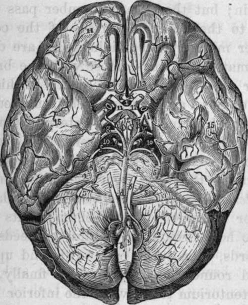
The Posterior Artery of the cerebrum is much larger than the superior artery of the cerebellum: at its origin the third nerve hooks round it. It first proceeds forwards and outwards, then turns backwards and upwards, so as to wind round the crus cerebri: finally, it passes above the tentorium to arrive at the inferior surface of the posterior lobe of the cerebrum, to which it sends numerous branches which first ramify in the pia mater and afterwards penetrate the substance of the brain: immediately after its origin it gives off several small twigs, some of which pass through the locus perforatus into the third ventricle, while others are distributed on the crura cerebri, corpora albicantia, and tuber cinereum. Where it begins to curve backwards it receives the posterior communicating branch of the internal carotid; immediately afterwards it gives off a choroid branch, which curves round the crus cerebelli, and supplies the choroid plexus, velum interpositum, and tubercula quadrigemina. Lastly, it gives off a small but constant branch that supplies the fascia dentata.
We may now review the arteries which form what is called the Circle of Willis:—in front we have the anterior communicating artery; posterior and external to this, the anterior arteries of the cerebrum, then the trunks of the internal carotids; behind these the posterior communicating arteries; next the posterior arteries of the cerebrum; and most posteriorly the anterior termination of the basilar artery itself: it is, in fact, more a heptagon than a circle. Within the circle of Willis the following parts are embraced, viz., anteriorly the commissure of the optic nerves, and lamina cinerea; behind this the tuber cinereum and base of the infundibulum, then the corpora mammillaria, middle locus perforatus, and generally, though situated above the area of the circle, some of the filaments of the origin of the third pair of nerves.

Fig. 21. Arteries at the base of the Brain, Circle of Willis.
1, 1, Posterior Lobes of the Brain. 2, 2, Hemispheres of the Cerebellum. 3, 3, Flocculi or Pneumogastric Lobes. 4, 4, Lower surface of the Anterior Lobe of the Cerebellum. 5, 5. Trifacial or fifth pair of Nerves. 6, 6, Sixth pair of Nerves. 7, Portio Dura of the seventh pair. 8, Auditory Nerve or Portio Mollis of the seventh pair. 9,9, Third pair of nerves. 10, 10, Crura Cerebri. 11, 11, Optic Nerves and Commissure. 12, Tuber Cinereuin, Infundibulum, and Corpora Mammillaria. 13, 13, The Olfactory Lobes. 14, 14, Anterior Cerebral Lobes. 15, 15, The Middle Lobes of the Brain, a, a, Vertebral Arteries, b, b, Anterior Spinal Arteries before their union, c, c, Inferior Arteries of the Cerebellum, at one side arising from the Basilar trunk, at the opposite side from the Vertebral, d, d, Basilar Artery, e, e, Anterior Arteries of the Cerebellum, f, f, Superior Arteries of the Cerebellum, g, g, Posterior Arteries of the Cerebrum, h, h, Posterior Communicating Arteries from the Internal Carotid, i, i, Internal Carotid Arteries, k, k, Anterior Cerebral Arteries connected by the Anterior communicating branch, on which is situated the Ganglion of Ribes. l, Anterior communicating Artery.
It may be remarked that where the vertebral artery ascends through vertebrae which have but little motion between each other, it is not tortuous; but in the superior part of the neck it makes a double curve,—first between the axis and atlas, and then between the atlas and occipital bone, in order as it were to escape injury; for in this manner, in passing from one of these bones to the other, it traverses twice the length of their vertical distance from each other; so that, as Mr. Mayo observes, the artery is only unbent, not stretched, in the more extensive motions of these bones. The vertebral artery has been known to be torn in fractures through the base of the skull.
The next branches of the subclavian artery are the internal mammary and thyroid axis, both of which arise opposite the internal margin of the scalenus anticus muscle, the former from the lower, and the latter from the upper and anterior surface of the artery.
Continue to:
