The Anterior Circumflex Artery
Description
This section is from the book "Anatomy Of The Arteries Of The Human Body", by John Hatch Power. Also available from Amazon: Anatomy of the Arteries of the Human Body, with the Descriptive Anatomy of the Heart.
The Anterior Circumflex Artery
The Anterior Circumflex Artery is a very small but very constant branch : it passes horizontally forwards and outwards, covered by the coraco-brachialis muscle and short head of the biceps. It then crosses the bicipital groove, covered by its synovial membrane and by the long head of the biceps, and sinks beneath the deltoid muscle, in the substance of which it anastomoses with the posterior circumflex artery. The anterior circumflex artery supplies the coraco-brachialis, biceps, and sub-scapularis muscles. While crossing behind the long head of the biceps, it sends a delicate branch upwards along the bicipital groove, to supply the head of the humerus and capsular ligament of the shoulder-joint.
It will be useful in the present stage of the dissection to take a glance at the principal arteries which form what is termed the scapular anastomosis. By means of this free arterial communication around the scapula, the blood of the subclavian artery will readily find its way into the arm or fore-arm in cases where the subclavian has been tied in its second or third stages, or where the axillary artery has been tied in its first or second stages. Along the axillary margin of the scapula we observe the continued branch of the sub-scapular artery passing towards the inferior angle of that bone: along the posterior or vertebral margin we see the posterior scapular passing towards the same point; and in relation with the superior or coracoid margin we find the supra-scapular artery. At the inferior angle of the scapula a free communication exists between the posterior scapular and sub-scapular arteries; at the posterior superior angle a similar communication exists between the posterior and supra-scapular arteries; and at the glenoid angle, underneath the root of the acromial process, a free anastomosis takes place between the supra-scapular and the sub-scapular arteries. Thus the axillary and subclavian arteries communicate freely with each other.
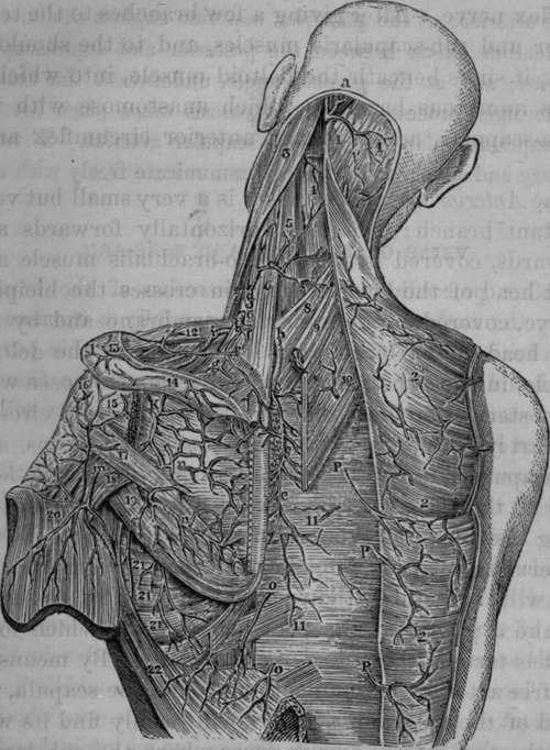
Fig. 26. Represents the Arteries of the Posterior part of the Neck and Shoulder. The Scapular Anastomosis.
1, 1, Occipital portion of Trapezius Muscle of each side. 2, 2, 1, Arterial branches to the Trapezius and Latissimus Dorsi Muscles. 3, Sterno-cleido-mastoid Muscle. 4, Splenius Capitis. 5, Splenius Colli. 6, 6, Levator Anguli Scapulae. 7, Lower portion or Sterno-mastoid. 8, Serratus Posticus Superior. 9. Rhomboideus Minor. 10, Rhomboideus Major divided. 11, 11, Aponeurosis covering long Muscles of Rack. 1*2, Clavicle with Arterial twig. 13, 14. Spine of Scapula with Arterial Twigs. 15, Insertion of Infraspinatus Muscle. 16, Capsule of shoulder-joint. 17, Teres Minor. 18, Long Head of Triceps between the two Teres Muscles. 19, Teres Major. 20, Deltoid divided and turned downwards. 21, 21 21, Serratus Magnus with Arterial Twigs. 22, Latissimus Dorsi divided and turned over. a. Occipital Artery emerging from underneath the Splenius Muscle to get into its third stage, b, b, Posterior Scapular Artery, c, c, c, Terminating branch of the Posterior Scapular Artery, d, e, Cervicalis Superticialis Artery cut. f, Twig to the Clavicle, g Small branch to Supra-spinatus. i, Supra-scapular Artery, k, Infra-Spinata Artery. 1, Acromion Process, m. Posterior Circumflex Artery, n, n, Anastomoses between the Infra-spinata, the Posterior branch of Subscapular and Posterior Scapular Arteries, o, o, Branches of the Intercostal Arteries. P, P, P, Dorsal branches of the Intercostal Arteries.
Continue to: