Deep Palmar Arch
Description
This section is from the book "Anatomy Of The Arteries Of The Human Body", by John Hatch Power. Also available from Amazon: Anatomy of the Arteries of the Human Body, with the Descriptive Anatomy of the Heart.
Deep Palmar Arch
The Deep palmar arch of arteries is covered in front by all the nerves, tendons, and muscles of the palm of the hand except by the interosseous muscles, which, together with the metacarpal bones, lie behind it. It crosses these bones nearly at right angles, lying close to their carpal extremities, and forming a slight curvature, the convexity of which looks towards the phalanges. This arch is accompanied by a branch of the ulnar nerve, which passes in company with the communieans profunda branch of the ulnar artery into this deep-seated situation of the hand: the nerve lies on the anterior surface of the arch and terminates in the muscles of the thumb. The deep palmar arch gives off the following branches:—

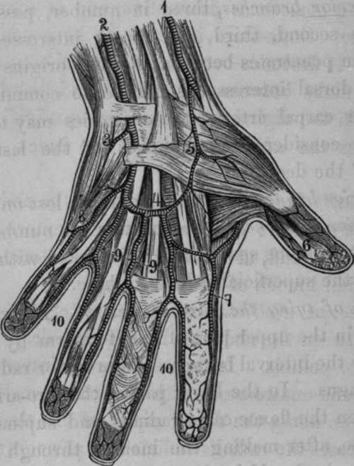
Fig. 32. Arteries of the Hand; Palmar Surface.
1. Radial Artery. 2, Ulnar. 3, Communicating Branch with the Deep Palmar Arch. 4, Superficial Palmar Arch. 5, Superficial Volar Artery. 6, Digital Arteries of the Thumb. 7, Radial Index Artery. 8, Digital Artery to the Little Finger. 9, Common Digital Arteries, 10, Digitals to the Fingers.
Anterior. Superior.
Posterior. Inferior.
The anterior branches are small, and are lost in the lumbri-cales muscles.
The posterior branches, three in number, pass backwards towards the second, third, and fourth interosseous spaces: each of them penetrates between the two origins of the corresponding dorsal interosseous muscle, to communicate with the posterior carpal artery: these arteries may therefore be indifferently considered as branches of the last-mentioned artery, or of the deep arch.
The superior branches are small, and are lost on the carpus.
The inferior branches, three or four in number, descend along the interosseous spaces, and anastomose with the digital branches of the superficial palmar arch.
Continue to:
