The Pleura And Lungs
Description
This section is from the book "Landmarks And Surface Markings Of The Human Body", by Louis Bathe Rawling. Also available from Amazon: Landmarks and Surface Markings of the Human Body.
The Pleura And Lungs
The Pleural Sacs
When the shoulders are depressed, (Fig. XVI, 5.) the two clavicles lie practically at right angles to the long axis of the body, (Fig. XVII, 4.) and in this position the apices of the pleural sacs extend into the supraclavicular region, lying about 1 1/2 inches above the clavicle under cover of the clavicular head of the sterno-cleido-mastoid muscle.
The anterior margin of each sac sweeps downwards and inwards behind the corresponding sterno-clavicular joint, the two sacs converging towards the angle of Ludwig, at which level they meet one another just to the left of the middle line. They then pass vertically downwards parallel to one another as far as the level of the fourth chondro-sternal junction.
The right sac passes onwards in the same straight line to the sixth or seventh chondro-sternal articulation, and then sweeps round the anterior, lateral, and posterior aspects of the chest wall, cutting across
(1) The upper part of the eighth costal cartilage in the lateral vertical line;
(2) The tenth rib in the mid-axillary line ;
(3) The eleventh rib in the line of the inferior angle of the scapula;
(4) The twelfth rib at the outer border of the erector spinae muscle.
The obliquity of the twelfth rib causes the pleura to fall below the level of the inner half of the rib, the pleura in this last part of its course being directed inwards towards the spine of the twelfth dorsal vertebra.
The left pleura, from the level of the fourth left chondro-sternal articulation, sweeps obliquely outwards and downwards behind the costal cartilages of the fifth, sixth and seventh ribs to the eighth costal cartilage in the lateral vertical plane. Beyond this point the left pleura follows the same general direction as the right sac, descending, however, to a slightly lower level.
The lowest limit reached by the two pleural sacs is situated in the mid-axillary line, the sacs there cutting across the tenth rib about 2 inches above the costal margin, which is in this situation usually represented by the tip of the eleventh rib.
Difficulty is sometimes experienced in endeavouring to verify the position of the twelfth dorsal spine, and in such cases the pleural reflection behind the mid-axillary region may be represented by a line which passes almost transversely backwards toward the median posterior line from the tenth rib in the mid-axillary line.
The two sacs are for the most part separated from one another by the width of the vertebral bodies (Fig. XVII, 4.) about 1 1/2 inches. In the lower part of the posterior mediastinum, however, the right sac approaches the median line.
2. The lungs may be mapped out in part by lines similar to those given for the pleural sacs. The apex of the upper lobe of each lung extends into the supra-clavicular region, (Fig. XVI, 6., Fig. XVII, 5.) and the anterior margins converge towards the angle of Ludwig, cutting obliquely across the corresponding sterno-clavicular joint.
The two anterior borders do not, however, meet at the angle of Ludwig, for the right lung on reaching the middle line passes vertically downwards as far as the level of the sixth or seventh chondro-sternal junction, whilst the left lung runs down behind the left border of the sternum to the junction of the fourth costal cartilage with the sternum.
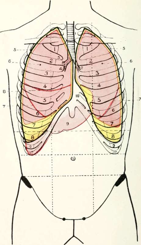
Fig. XVI. The Pleural Sacs, Lungs, Etc
1. | The trachea. | 7. | The main oblique fissure of the lungs. |
2. | The left bronchus. | ||
3. | The eparterial bronchus. | 8. | The small transverse fissure or the right lung. |
4. | The hyparterial bronchus. | ||
5. | The pleurae. | 9. | The liver. |
6. | The lungs. | 10. | Area of superficial cardiac dull-ness. |
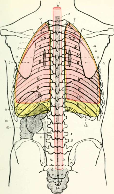
Fig. XVII. The Pleural Sacs, Lungs, Etc
1. | The spinal dural sheath. | 9. | The spleen. |
2. | The spinal cord. | 10. | The left kidney in Morris's quadrilateral. |
3. | The filum terminale. | ||
4. | The pleurae. | 11. | The ureter. |
5. | The lungs. | 12. | The liver. |
6. | The main oblique fissures of the lungs. | 13. | The descending colon. |
14. | The rectum. | ||
7. | The apex of the lower lobe of the lungs. | 15. | The posterior superior iliac spine. |
8. | The roots of the lungs. |
* The transverse processes of the first and second lumbar vertebra; should be drawn in such a manner as to come into contact with the inner border of the kidney.
The right lung, from the level of the sixth or seventh chondro-sternal junction,sweeps outwards, cutting across
(1) The sixth costal cartilage in the lateral vertical line
(2) The eighth rib in the mid-axillary line ;
(3) The tenth rib in the line of the inferior angle of the scapula, and finally passing inwards towards the tenth dorsal spine.
The left lung, from the outer border of the sternum at the level of the fourth chondro-sternal articulation, passes outwards for a short distance along the lower border of the fourth costal cartilage, and then turns downwards and inwards in a curved direction to the sixth costal cartilage in the lateral vertical plane. The lung then sweeps round the chest wall, following a course similar to that already indicated as pursued by the right lung, the left lung lying, however, at a slightly lower level.
Stress should, perhaps, be laid on the fact that, as the lower border of each lung sweeps round the antero-lateral, lateral, and posterior aspects of the chest wall, from the sixth costal cartilage in front to the tenth dorsal spine behind, the course pursued is practically transverse to the long axis of the body.
It will be convenient to here put in a tabulated manner the comparative lower levels of the right pleura and lung. Starting in each case at the right chondro-sternal junction, the pleura and lung may be represented by lines which traverse the chest wall, cutting across the
(1) Lateral vertical line at the eighth costal cartilage (pleura)sixth costal cartilage (lung).
(2) Mid-axillary line at the tenth rib (pleura)eighth rib (lung).
(3) Scapular line at the eleventh rib (pleura)tenth rib (lung).
(4) And sweeping in towards the twelfth dorsal spine (pleura)tenth dorsal spine (lung). The numbers to be remembered are, therefore, 6, 8, 10, 11, and 12 for the pleura, and 6, 6, 8, 10, 10 for the lung.
On the left side, the levels are similar with two main exceptions(a) the lung and pleura sweep outwards so as to leave a part of the heart uncovered (see " superficial cardiac dulness"), (b) both lung and pleura descend to a slightly lower level.
The Lower Limit of the Lung, the Pleura, and the Liver in the Right Mid-axillary Line.
(1) The lung corresponds to the eighth rib.
(2) ,, pleura tenth rib.
(3) liver costal margin, or even lower.
Continue to: