Tenth Or Vagus Nerve
Description
This section is from the book "Nerves Of The Human Body", by Charles R. Whittaker. Also available from Amazon: Hughes Nerves Of The Human Body.
Tenth Or Vagus Nerve
Deep Origin
Similar to that of the glossopharyngeal nerve, but only a few afferent fibres pass to the tractus solitarius.
Superficial Origin
Twelve to fifteen rootlets emerge from the medulla, lateral to the restiform body, and between the glossopharyngeal above, and the spinal accessory below.
Course
The nerve leaves the cranium through the posterior compartment of the jugular foramen. It occupies the same sheath of dura mater as the spinal accessory. As the vagus lies in the foramen, two ganglia are formed in connection with it. The upper and smaller one is termed the ganglion jugulare, while the other is called the ganglion nodosum.
The nerve descends between the internal carotid artery and the internal jugular vein. Entering the carotid sheath the vagus travels downwards, lying behind and between the common carotid artery and internal jugular vein.
The Right Vagus passes over the first part of the subclavian artery, and reaches the thorax behind the right innominate vein. In the superior mediastinum it is found on the right side of the innominate artery and trachea, and is posterior to the superior vena cava. After running along the lateral margin of the trachea to the posterior mediastinum, the nerve splits up into several branches at the back of the root of the lung; these form the posterior pulmonary plexus. From the plexus the vagus issues as two cords, which after crossing the vena azygos major, pass on to the oesophagus and unite with the vagus of the opposite side as the oesophageal plexus. The nerve leaves the plexus as a single trunk which descends in front of the gullet, traverses the corresponding opening in the diaphragm, and is distributed to the posterior surface of the stomach. Communicating fibres are furnished to the coeliac, splenic, and left renal plexuses.
The Left Vagus enters the thoracic cavity between the left common carotid and subclavian arteries, lying posterior to the left innominate vein and left phrenic nerve. After crossing in front of the aortic arch, it breaks up at the back of the root of the lung into the posterior pulmonary plexus. From this plexus the two efferent nerves travel over the descending thoracic aorta, and reaching the oesophagus, form the oesophageal plexus. The issuing nerve passes through the oesophageal orifice of the diaphragm in front of the gullet. It ramifies over the anterior surface of the stomach; certain of the branches pass in the gastro-hepatic ligament to the hepatic plexus.
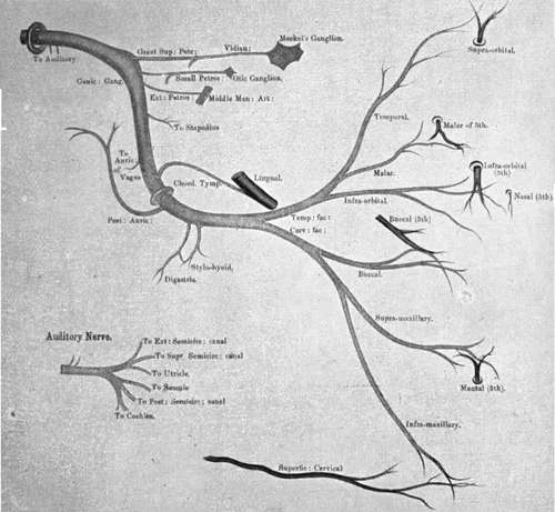
Plate IV. Facial Nerve
Branches
(A) Meningeal from the jugular ganglion innervates the dura mater of the posterior fossa.
(B) Auricular (Arnold's nerve) arises from the jugular ganglion, and receives a small twig from the glossopharyngeal. It enters the skull through a minute orifice in the jugular fossa, traverses the petrous part of the temporal, where it communicates with the facial, and emerges from the bone at the auricular fissure. Arnold's nerve supplies the skin of the posterior aspect of the auricle, communicating there with the posterior auricular.
(C) Pharyngeal springs from the upper part of the ganglion nodosum. Its fibres are derived from the spinal accessory. This branch reaches the pharyngeal wall by passing between the external and internal carotid arteries. It divides into several branches which join branches of the glossopharyngeal and cervical sympathetic to form the pharyngeal plexus. The plexus distributes twigs to the pharyngeal mucous membrane, constrictors of the pharynx, and all the muscles of the soft palate, with the exception of the tensor palati (supplied by the otic ganglion). One branch (the lingual) from the plexus communicates with the hypoglossal. {d) Superior laryngeal takes origin from the ganglion nodosum about its middle. It passes downwards and medially behind the external and internal carotid arteries. The nerve bifurcates into a small lateral branch, the external laryngeal, which supplies the inferior constrictor and crico-thyreoid muscles, and a larger internal laryngeal. The latter insinuates itself between the middle and inferior constrictors to perforate the thyreo-hyoid membrane. It innervates the laryngeal mucous membrane, and communicates with the recurrent (inferior) laryngeal nerve. The motor fibres of the superior laryngeal come from the spinal accessory,
(E) Recurrent (Inferior) Laryngeal
On the right side this nerve arises in the lower part of the neck, and hooks backwards beneath the first part of the subclavian artery. The left recurrent laryngeal takes origin in the thorax, and winds backwards around the aortic arch on the lateral side of the ligamentum arteriosum. Each nerve passes obliquely upwards and medially behind the subclavian, common carotid, and inferior thyreoid arteries. In addition, on the left side the nerve occupies the groove between the oesophagus and trachea. It is sheltered by the inferior pole of the thyreoid body, and reaches the larynx by passing beneath the inferior constrictor muscle. The recurrent laryngeal supplies the intrinsic muscles of the larynx, the fibres being derived from the spinal accessory. In addition to communicating with the inferior cervical ganglion of the sympathetic and with the internal laryngeal, the recurrent laryngeal furnishes branches to the heart, trachea, oesophagus, and inferior constrictor.
(F) Cardiac
There are two groups, cervical and thoracic.
(1) Cervical; the upper ones are of small size and join the deep cardiac plexus. The lower branch which arises at the root of the neck, differs on the two sides. That on the right side passes to the deep cardiac plexus, while the left one crosses in front of the aortic arch to enter the superficial cardiac plexus.
(2) Thoracic; these branches arise in the superior mediastinum. On the right side they are partly derived, while on the left side they are entirely derived, from the recurrent laryngeal. In both cases they pass to the deep cardiac plexus. (g) Pulmonary.-A few twigs reach the front of the root of the lung, which join with branches of the cardiac plexuses (see sympathetic nerves) to join the anterior pulmonary plexus. The posterior pulmonary plexus is composed of the greater part of the vagus, together with branches from the second, third, and fourth thoracic sympathetic ganglia. From the plexuses twigs accompany the bronchi and pulmonary vessels to the lungs.
(H) Oesophageal
These branches are given off above and below the root of the lung.
Communications
In addition to the communications already mentioned, the jugular ganglion receives twigs from the superior cervical ganglion of the sympathetic, and the spinal accessory. The nodal ganglion communicates with (a) the superior cervical ganglion of the sympathetic; (b) the hypoglossal; (c) the loop between the first and second cervical nerves; (d) the spinal accessory. It is by means of the last-named communication that the vagus nerve receives inhibitory fibres for the heart, together with the motor fibres for the soft palate, oesophagus, larynx, stomach, intestines, and lungs.
Continue to: