85. The Heart In Mammals
Description
This section is from the book "Animal Physiology: The Structure And Functions Of The Human Body", by John Cleland. Also available from Amazon: Animal Physiology, the Structure and Functions of the Human Body.
85. The Heart In Mammals
The Heart In Mammals, as will be seen from what has been said, is divisible into a right and a left part, each of which is comparable with a fish heart, consisting, as it does, of an auricle and a ventricle; these parts are completely separate, one from the other, from the time of birth, so far as the blood contained in them is concerned; but they act synchronously, and are structurally one heart. Anatomically considered, the natural division of the heart is into an auricular and ventricular part, separated by a deep sulcus, the auriculo-ventricular groove. The ventricular part is a strong muscular structure invested completely with the serous covering of the pericardium, and unconnected with other viscera. It is directed downwards, forwards, and to the left side, resting on the diaphragm in man, and narrowing to the apex, which is felt beating opposite the interval between the sixth and seventh costal cartilages of the left side. The apex is formed entirely by the walls of the left ventricle, which are three times as thick as those of the right ventricle, the blood requiring much greater force to propel it through the system than to send it through the lungs; and if the ventricles be cut across, the section of the left ventricle will be seen to be circular, while the right is curved crescentically round it.
Above the auriculo-ventricular groove, ascending from the base of the ventricles, the two arterial trunks issuing from those two cavities lie close together behind the breast-bone, each twisted somewhat round the other. That which rises from the right ventricle is the pulmonary artery, and divides into a right and left branch, one going to each lung; while the systemic artery, arising from the left ventricle, and concealed at its origin by the pulmonary artery, is called the aorta.
The auricles have exceedingly thin muscular walls, their whole function being to receive the blood, which continues pouring in during the contraction of the ventricles, and to pass it into them through the large auriculo-ventricular apertures as soon as they relax. They lie behind and to the sides of the arterial trunks, and each is prolonged into a pointed cul-de-sac in front, which, from a fancied resemblance to a dog's ear, is called the auricular appendage; and these appendages have given their name of auricles to the cavities to which they belong. The cavities are separated one from the other by a thin septum, which, as seen from the right auricle, presents a depression and, in front of it, a crescentic border, the fossa and annulus ovalis, marking the position of an opening which exists, and is made use of, in foetal life, but is shut up after birth. In the rare instances in which it continues after birth to allow blood to pass through it, the circulation of dark blood in the system is the result, constituting the disease called cyanosis, and destroying life.
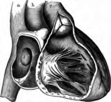
Fig. 65. Right Side of the Heart, a, b, Superior and inferior vena cava entering the right auricle; c, Eustachian valve; d, annulus ovalis; e,f, anterior and posterior cusps of the tricuspid valve descending from the margin of the auriculo-ventricular opening; g, pulmonary artery with its orifice shut by distension of the three, pouches of its semilunar valve; h, aorta.
The right auricle receives blood in two streams nearly vertically opposite one another, one from the vena cava superior, bringing the blood from the head and upper limbs, the other from the vena cava inferior, bringing the blood from the lower limbs and greater part of the trunk; while, in addition, the blood from the walls of the heart enters by one considerable and several smaller orifices. The left auricle receives its blood by streams transversely opposite one another, entering by the pulmonary veins from the right and left lungs.
86. The heart can be seen in action by laying open a frog, but still more satisfactorily in the chest of a mammal. The auricles are seen to contract first, and to distend the ventricles with blood; the ventricles contract immediately afterwards, and then there is a pause before the auricles become quite distended and contract again. The contraction of the auricles is completed in about a third of the time taken by the ventricles to contract; but it is not thorough, for it proceeds in a wave forwards from the venous trunks to the tips of the appendages, so that the appendages are at first distended, and when in turn they contract, the rest of the auricular walls are already relaxed. On account of this mode of contraction of the auricles, there is in health little tendency of the blood to regurgitate into the venous trunks; and the mouths of these vessels are unguarded in mammals, although protected by competent valves in other animals. There is in man a fold of membrane, the Eustachian valve, in front of the vena cava inferior; but it can have little action as a valve after birth, for it is frequently nearly absent in the adult. The ventricles contract in a different way from the auricles. The muscular fibres in the middle depth of their walls embrace them circularly, while the successively deeper and more superficial layers have successively steeper degrees of obliquity, and are continuous one with another both at base and apex; and in consequence of this arrangement, these cavities are contracted throughout their whole extent by both shortening and narrowing at the same time, till they are completely emptied. But no matter how completely or forcibly the ventricles might contract, they would make but an inefficient engine of propulsion were there not some means of preventing the blood being pushed, during their contraction, back into the auricles, and recoiling, after their contraction, back into them from the arteries. Such waste of power is prevented by the presence of valves, which guard the arterial and auriculo-ventricular orifices.
Continue to: