The Veins Which Accompany The Spermatic Artery
Description
This section is from the book "Anatomy Of The Arteries Of The Human Body", by John Hatch Power. Also available from Amazon: Anatomy of the Arteries of the Human Body, with the Descriptive Anatomy of the Heart.
The Veins Which Accompany The Spermatic Artery
The Veins Which Accompany The Spermatic Artery arise from the testis and epididymis, and form a plexus immediately after their junction. They then ascend, four or five in number, through the inguinal canal, lying in front of the vas deferens, and ultimately unite, in the lower part of the lumbar region, into a single trunk which ascends on the outside of the spermatic artery. The right spermatic vein empties its contents into the inferior vena cava, with which it forms an acute angle below the termination of the right renal. The left spermatic vein empties itself into the left renal, with which it forms nearly a right angle. In many cases the spermatic vein divides at a short distance above the gland, into many branches, so as to form a peculiar plexus, termed the pampiniform plexus; after which it again becomes a single trunk. Meckel says that the plexus exists more frequently on the left side than on the right.
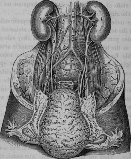
Fig. 42. Represents the Arteries of the, Uterus in a Female who died six days after delivery.
A, Abdominal Aorta. B, Superior Mesenteric Artery, divided. C, C, Renal Arteries. D, Inferior Mesenteric Artery, cut. E, E, Common Iliac Arteries. F, F, External Iliac Arteries. G, G, Internal Iliac Arteries, a, a, a, Spermatic Arteries, greatly convoluted and enlarged at their termination, b, b, b, b, b, Lumbar Arteries, c. Middle Sacral Artery, d, d, d, d, Uterine Arteries, convoluted and enlarged, e, e, Internal Circumflexae Ilii Arteries, f. f, Anastomosis between Iliolumbar and Circumflexae Ilii Arteries. 1, 1, Kidneys. 2, 2t Pelves of the Kidneys. 3, 3, Quadratus Lumborum Muscle of each side. 4, 4, Psoas Parvus. 5, 5, Psoas Magnus. 6, 6, Iliacus Internus. 7, 7, Anterior Superior Iliac Spine. 8, 8, Crural Arch. 9, Promontory of Sacrum. 10, Rectum. 11, Uterus turned forward. 12, 12, The ovaries. 13, 13, The Fallopian Tubes.
As the left testicle is lower than the right, the veins are longer; and this (together with the peculiar mode of termination of the left spermatic vein, and its relation to the sigmoid flexure of the colon) is supposed to explain why a varicose state of these vessels is more frequent on the left than on the right side.
In the male foetus these arteries are proportionably very short, as the testicles are placed within the abdomen during the greater part of intra-uterine life; but they subsequently elongate as the testicles descend. The spermatic arteries are remarkable for increasing in diameter as they recede from their origin. In the operations of castration, the spermatic artery is apt to contract considerably on being divided, so as to render it difficult to secure it in a ligature. The surgeon may avoid this embarrassment by holding the divided cord in his hand till an assistant draws out and secures whatever branches are necessary. The exquisitely painful practice of including the cord in the ligature is now universally abandoned.
Continue to: