Colica Sinistra. Arteria Sigmoidea; And Superior Haemorrhoidal
Description
This section is from the book "Anatomy Of The Arteries Of The Human Body", by John Hatch Power. Also available from Amazon: Anatomy of the Arteries of the Human Body, with the Descriptive Anatomy of the Heart.
Colica Sinistra. Arteria Sigmoidea; And Superior Haemorrhoidal
The Colica Sinistra ascends between the layers of the left mesocolon, and divides into a superior and inferior branch; the former anastomoses with the colica media, and the latter with the sigmoid artery.
The Sigmoid artery crosses the front of the psoas muscle, and divides into a superior branch, which communicates with the colica sinistra, and an inferior branch, which terminates in supplying the sigmoid flexure of the colon, and in anastomosing with the superior haemorrhoidal. This artery also supplies the psoas and iliacus muscles and the ureter.
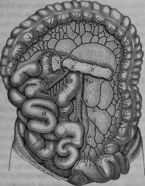
Fig. 38. Distribution of the Inferior Mesenteric Artery.
1, Aorta. 2, Inferior Mesenteric Artery. 3. Left Colic Artery. 4, Sigmoid Artery. 5, Superior Ilactnorrhoidul Artery. 6, Superior Mesenteric Artery. 7, Middle Colic Artery anastomosing with the left and the right (8) Colic Arteries. 9. Branches to the small Intestine. 10, Left Renal Artery, a, Small Intestine turned to the right side, b, Large Intestine, c, Pancreas.
The Superior Hsemorrlioidal artery cannot well be examined until the arteries of the pelvis have been dissected. If we suppose the rectum divided into three stages,a superior, middle, and inferior,we find that in the superior stage it is surrounded with peritoneum, and has a meso-rectum : in the middle stage it has no meso-rectum, but is covered by peritoneum upon its anterior part, and on a portion of its sides: in the inferior stage it has no peritoneal covering. Now, we find the artery distributed in conformity with this arrangement; for in the first of these stages it descends as a single trunk between the layers of the meso-rectum; it then divides, about four inches from the anus, into two branches; and these, in the second stage, follow (one on either side) the line of reflection of the peritoneum from the side of the rectum; lastly, in the third stage the terminating branches of the artery are numerous, and distributed all round the inferior extremity of the intestine. The superior haemorrhoidal artery communicates freely with the haemorrhoidal branches of the internal iliac and pudic arteries.
In case of hemorrhage into the rectum from the haemorrhoidal arteries, a membranous tube closed at one end may be introduced into the rectum, and through the other cold water may be injected with a syringe, so as to distend it, and thus compress the bleeding vessels on the surface of the gut. The water can be occasionally renewed without withdrawing the tube. Or, which is preferable, as in the case of hemorrhage after the operations for fistula in ano, or after the excision of haemorrhoidal tumors, a small fine linen bag, open at one end, and provided with tapes, may be introduced into the rectum, and through the external or open extremity a quantity of charpie may be introduced, so as to distend the bag; the tapes may be then tied across the stuffing of charpie, and the dressing secured: the necessary compression on the bleeding vessels of the intestines will be thus effected.
The liver, stomach, spleen, and intestines may now be examined and removed, after which the student may proceed with the dissection of the deeper arteries within the cavity of the abdomen.
Continue to: