Chapter VII. Benign Tumours
Description
This section is from the book "Cancer And Other Tumours Of The Stomach", by Samuel Fenwick. Also available from Amazon: Cancer and other tumours of the stomach.
Chapter VII. Benign Tumours
The benign tumours of the stomach and duodenum which have not been described in the previous chapters are myomata, fibromata, lipomata, lymphadenomata, mryxomata, osteomata, and aneurysms. They are all very rare, and, with perhaps the exception of the first, are practically devoid of clinical interest.
(1) Myoma
This was first described in 1762 by Morgagni, since which time more than forty other cases have been recorded. As a rule it takes the form of an oval or round, firm, solitary tumour situated near the cardiac orifice or at the greater curvature, but it is sometimes encountered in the pyloric end of the stomach or in the duodenum (Wesener). In most cases it does not exceed the size of a pea or a cherry, and occupies the substance of the gastric wall; but occasionally it forms an enormous lobulated or knotty tumour, which projects into the abdominal cavity beneath the peritoneum. On section the growth is found to have originated in the muscular coat, and presents a brown or milk-white colour, and on microscopical examination it is seen to consist of interlacing bundles of unstriped muscle, mixed with strands of fibrous tissue and arranged in concentric layers. The tumour is of slow growth and is usually met with in men of middle age. The submucous variety is prone to undergo cystic degeneration and to become pedunculated, while the subserous form occasionally assumes a sarcomatous character (Brodowski). Kidd reported a submucous fibro-myoma, two inches in length and three-quarters of an inch in width, which involved the cardiac orifice, and Vogel one about the size of an almond which was situated near the lower end of the oesophagus. V. Erlach is said to have removed a myoma from the stomach which weighed 5,400 grammes, v. Eiselsberg one about the size of a man's head, and Kunze a lipomyoma '251 grammes in weight.
Symptoms
Small myomata are not accompanied by any special symptoms unless the mucous membrane which covers them has undergone ulceration or they happen to obstruct one of the orifices of the stomach. In the former case the patient may suffer from pain after food and repeated attacks of hasmatemesis, while in the latter there is dysphagia or periodic vomiting. Herhold has recorded an example of myoma in a woman thirty-seven years of age who for three years had suffered from severe vomiting after meals. The stomach was dilated and the vomit contained free hydrochloric acid. An exploratory operation showed the existence of a myoma about the size of a hazel-nut, which had produced obstruction of the pylorus. The duodenal myoma described by Wesener was accompanied by vomiting and emaciation, and the signs of dilatation of the stomach, which caused it to be mistaken for cancer of the pylorus.
The subject of Kunze's case was a man aged fifty-two, who had suffered from abdominal pain for fourteen years. There was no vomiting or cachexia, but he was extremely emaciated. On examination a tumour about the size of a man's fist could be felt close to the navel, the existence of which had been known to the patient for four years. It was hard, nodular and painless, slightly movable, and sub-tympanitic on percussion. No details are given of v. Erlach's case, but since an operation was undertaken and the tumour was of enormous size it must have been accompanied by symptoms of sufficient severity to necessitate active treatment.
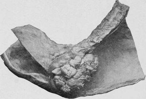
Pig. 68.-Papilloma of the pylorus. (Museum of the Royal College of Surgeons.).
Continue to:
- prev: Case XLVI
- Table of Contents
- next: Fibromata