Choroid
Description
This section is from the book "Wonders Of The Human Body", by Auguste Le Pileur. Also available from Amazon: Wonders of the Human Body.
Choroid
On the internal surface of the sclerotic is a vascular membrane called the choroid, which lines it closely from the bottom of the eye to the circumference of the cornea, and is attached to it by a very fine cellular tissue. The choroid is composed of two layers, of which the external corresponds to the sclerotic, and the internal, or membrane of Ruysch, corresponds to the retina. These two layers, attached to each other by their internal surfaces, are covered externally with a layer of pigment, which is thicker next to the retina than on the side toward the sclerotic. The choroid is pierced behind by an opening, which gives passage to the optic nerve; in front, near the circumference of the cornea, it separates in order to form the ciliary circle and ciliary processes. The ciliary circle, ring, or muscle, is a little band, vascular, like the choroid, slightly adherent by its external surface to the sclerotic, and united by its lesser circumference to the cornea, at the point where the latter attaches itself to the sclerotic. Behind the ciliary circle a series of membranous rays are seen joined together, and forming a crown; these are the ciliary processes (from processus, prolongation, ray), the whole of which constitute the ciliary body or disk. These rays, which are attached to the choroid, like the ciliary circle, are of two kinds; one into which the crystalline is set, and which gives attachment to its capsule, termed the ciliary processes of the vitreous body; the others extend to the iris, behind which they form a sort of circular curtain, by folding back on themselves, and adhering to the larger circumference of this membrane. Thus fixed by one border, the ciliary disk floats by the other, like a fringe behind the iris, yielding to the slightest impulse which may be communicated to it. The ciliary processes are covered with a thick layer of pigment.
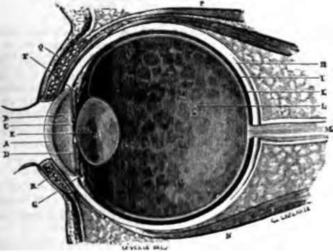
Fig. 34. Vertical section of the eye on the median line.
A. Cornea.
B. Anterior chamber.
C. Pupil.
D. Iris.
E. Crystalline.
F. Zone of Zinn, forming the anterior wall of canal of Petit.
G. Ciliary processes and circle.
H. Sclerotic.
I. Choroid. K. Retina. L. Vitreous body. M. Optic nerve. N. Right inferior muscle. O. Right superior muscle. P. Levator muscle of eyelid. Q. Lachrymal glands. K. Lachrymal canal.
Continue to:
- prev: Chapter XI. Sense Of Sight
- Table of Contents
- next: Iris