The Three Kinds Of Nerve Tissue
Description
This section is from the book "The Human Body: An Elementary Text-Book Of Anatomy, Physiology, And Hygiene", by H. Newell Martin. Also available from Amazon: The Human Body.
The Three Kinds Of Nerve Tissue
The microscope shows that the nervous organs contain tissues peculiar to themselves, known as nerve-fibres and nerve-cells. The cells are found in the centres only; while the fibres, of which there are two main varieties known as the white and the gray, are found in both trunks and centres; the white variety predominating in the cerebro spinal nerves and in the white substance of the centres, and the gray in the sympathetic trunks and the gray portions of the central organs.
How are the ganglia of the sympathetic system arranged?
How is each united to others? How far through the body do the chains of sympathetic ganglia extend? Where are the sympathetic chains situated in the trunk of the body?
How are the sympathetic ganglia united with spinal and cranial nerves? What arise from the ganglia? What do they form in the trunk region of the body? Where do many sympathetic nerve-fibres end? How do sympathetic nerves differ to the unaided eye from spinal or cerebral nerves? How is the brain enabled to control the parts supplied by the sympathetic system ?
How many kinds of nerve-tissue are there? What are the peculiar nerve-tissues called?
White Nerve Fibres
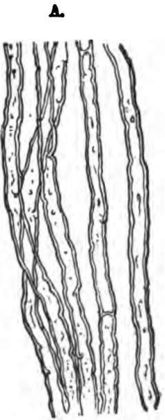
White Nerve Fibres consist of extremely delicate threads, about 1/2000 inch in diameter, but frequently of a length which is in proportion very great. Each is continuous from a nerve-centre to the region in which it ends, so that the fibres, e.g., which pass out from the spinal cord and run on to the skin of the toes, are three to four feet long. If a perfectly fresh white nerve-fibre be examined with the microscope it presents the appearance of a homogeneous glassy thread; but soon it acquires a characteristic double border (Fig. 85) from the coagulation of a portion of its substance, as a result of which three layers are brought into view. Outside is a thin transparent envelope (1, Fig. 86) called the primitive sheath; inside this is a fatty substance, 2, forming the medullary sheath (the coagulation of which gives the fibre its double border), and in the centre is a core, the axis cylinder, 3, which is the essential part of the fibre, since near its ending the primitive and medullary sheaths are frequently absent. At intervals of about 1/25 inch along the fibre are found nuclei. These are indications of the primitive cells which by their elongation, fusion, and other modifications have built up the nerve-fibre. In the course of a nerve-trunk its fibres rarely divide; when a branch is given off some fibres merely separate from the rest, much as a skein of silk might be separated at one end into smaller bundles containing fewer threads.

Fig. 85. White nerve-fibres soon after removal from the body and when they have acquired their double contour.

Fig. 86. Diagram illustrating the structure of a white or medullated nerve-fibre. 1,1, primitive sheath; 2, 2, medullary sheath; 3, axis cylinder.
Where only do we find nerve-cells? Where are nerve-fibres found? How many main kinds of nerve-fibres are there? Name them. Where do the white nerve-fibres predominate? Where the gray ?
Describe white nerve-fibres.
Gray Nerve Fibres
Gray Nerve Fibres have no medullary sheath, and consist merely of an axis cylinder and primitive sheath. Gray fibres are especially abundant in the sympathetic trunks; and they alone are found in the olfactory nerve.
Is each nerve-fibre continuous from centre to end? Point out nerve-fibres three or four feet long. What is the appearance under the microscope of a quite fresh white nerve-fibre? How does it soon alter? Name the layers then seen and state their relative positions. Which is the essential part of the nerve-fibre? Give a reason for your statement. What are found at intervals along the nerve-fibre? What do they indicate?
What occurs when a nerve-trunk branches?
Continue to:
