Chapter XIX. The Kidneys And The Skin
Description
This section is from the book "The Human Body: An Elementary Text-Book Of Anatomy, Physiology, And Hygiene", by H. Newell Martin. Also available from Amazon: The Human Body.
Chapter XIX. The Kidneys And The Skin
General Arrangement Of The Nitrogen Excreting Organs
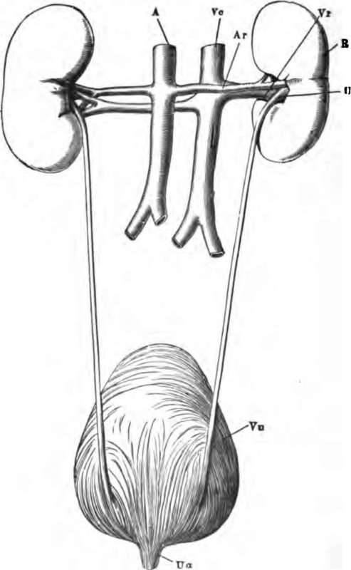
These organs are (1) the kidneys, the glands which secrete the urine; (2) the ureters or ducts of the kidneys, which carry the secretion to (3) the urinary bladder, a reservoir in which it accumulates and from which it is expelled from time to time through (4) an exit tube, the urethra. The general arrangement of these parts, as seen from behind, is shown in Fig. 73. The kidneys, R, lie at the back of the abdominal cavity, opposite the upper lumbar vertebrę, one on each side of the middle line. Each is a solid mass, with a convex outer and a concave inner border, and its upper end a little larger than the lower. From the abdominal aorta, A, a renal artery, Ar, enters the inner border of each kidney, to break up within it into finer branches, ultimately ending in capillaries. The blood is collected from these into the renal veins, Vr, one of which leaves each kidney and opens into the inferior vena cava, Vc, which carries it, after having lost water and urea in the kidney, back to the heart. From the concave border of each kidney proceeds also the ureter, U, a slender tube opening below into the bladder, Vu, near its lower end. From the bladder proceeds the urethra, at Ua. The channel of each ureter passes very obliquely through the wall of the bladder; accordingly if the pressure inside the latter organ rises above that of the liquid in the ureter the walls of the oblique passage are pressed together and it is closed. Usually the bladder (which contains muscular tissue in its walls) is relaxed, and the urine flows readily into it from the ureters. The commencement of the urethra being kept closed by elastic tissue around it (which can voluntarily be reinforced by muscles which compress the tube), the urine accumulates in the bladder. When this latter contracts and presses on its contents the ureters are closed in the way above indicated, the elasticity of the fibres closing the urethral exit from the bladder is overcome, and the liquid is forced out.
Name the chief organs concerned in removing from the body its nitrogenous waste matters. Describe the general arrangement of these organs. Describe the form of a kidney. What vessel supplies it with blood? What vessel carries blood out of the kidney? What has the blood carried oft from a kidney lost while flowing through that organ?

Fig. 73. The renal organs, viewed from behind. R, right kidney; A, aorta; Ar, right renal artery; Vc, inferior vena cava; Vr, right renal vein; U, right ureter; Vu, bladder; Ua, commencement of urethra.
Continue to:
