Naked Eye Structure Of The Kidneys
Description
This section is from the book "The Human Body: An Elementary Text-Book Of Anatomy, Physiology, And Hygiene", by H. Newell Martin. Also available from Amazon: The Human Body.
Naked Eye Structure Of The Kidneys
When a section is made through a kidney from its outer to its inner border (Fig. 74) it is seen that a deep fissure, the hilus, leads into the latter. In the hilus the ureter widens out to form the pelvis of the kidney, which breaks up into a number of smaller divisions, the cups or calices. The cut surface of the kidney proper is seen to consist of two distinct parts; an outer or cortical portion, and an inner or medullary. The medullary portion is less red and more glistening to the eye, is finely striated in a radial direction, and does not consist of one continuous mass, but of a number of conical portions, the pyramids of Malpighi, 2', each of which is separated from its neighbors by a prolongation,*, of the cortical substance. This, however, does not reach to the apex of the pyramid, which projects, as the papilla, into a calyx of the ureter. At its outer end each pyramid separates into smaller portions, 2", separated by thin layers of cortex and gradually spreading everywhere into the latter. The cortical substance is redder, more granular looking, and less shiny than the medullary; it forms everywhere the outer layer of the organ, besides dipping in between the pyramids in the manner above described.
What is the ureter? Under what circumstances is its opening into the bladder closed? What is the usual state of things? How is the commencement of the urethra ciosed? What results? What happens when the bladder contracts?
What is seen on a section made through a kidney? How is the pelvis of the kidney formed? What are the calices? What is seen, on the cut surface of the kidney proper? How does the medullary part differ in appearance from the cortical?
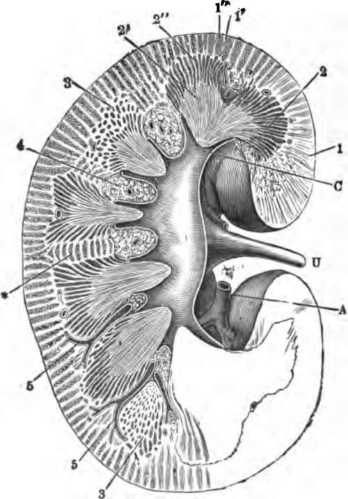
Fig. 74. Section through the right kidney from its outer to its inner border. 1, cortex; 2, medulla; 2', pyramid of Malpighi; 2", pyramid of Ferrein; 5, small branches of the renal artery entering between the pyramids; a, a branch of the renal artery; C, the pelvis of the kidney; U, ureter; C, a calyx.
Describe the pyramids of Malpighi What lies between them?
The renal artery divides in the hilus into branches (5) which run into the kidney substance between the pyramids, give off a few twigs to the pyramids, and end finally in a much closer vascular network in the cortex.
Continue to:
- prev: Chapter XIX. The Kidneys And The Skin
- Table of Contents
- next: The Minute Structure Of The Kidney