Chapter XI. Absorption
Description
This section is from the book "Animal Physiology: The Structure And Functions Of The Human Body", by John Cleland. Also available from Amazon: Animal Physiology, the Structure and Functions of the Human Body.
Chapter XI. Absorption
113. We have now to take into consideration the means by which the substance of the blood is replenished. This is effected by absorption, or the sucking up of material into vessels, partly from the alimentary canal, and partly from the tissues. Matter from both these sources is absorbed by the capillary blood-vessels, and so carried into the veins; but there is another set of vessels, the lymphatics, more especially referred to when absorbents are spoken of, whose whole office is one of absorption.
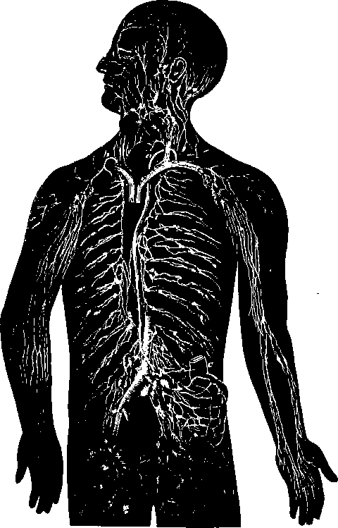
The lymphatics or absorbents are a system of vessels with delicate walls, and having the appearance of long and slender threads when they are empty. The trunk into which the majority of them pour their contents, the thoracic duct, is no greater in diameter than a small crow quill, and sometimes not so large. The thoracic duct begins in the upper part of the back of the abdomen, where it forms a dilatation four or five times its width in the rest of its course, named the receptaculum chyli, and runs up through the thorax in front of the vertebral column, to open, at the root of the neck, into the angle of junction of the left jugular and left subclavian veins. It receives the absorbents from the whole body, with the exception of the right half of the thorax, right arm, and right side of the head and neck. The absorbents from these parts unite to form a short trunk, which opens into the angle of junction of the right jugular and subclavian veins, and is called the right lymphatic duct prevent the injection of fluid backwards from the larger trunks into the radicles. These valves are set so thickly as to give to the vessels, when filled, a beaded appearance, there being a dilatation opposite each valvular pouch. In the limbs and in the walls of the trunk, they are arranged in a deep and superficial set; and in the viscera there is usually, in like manner, a set on the surface of an organ, and a deep set accompanying its blood-vessels.
The lymphatic vessels are difficult to study on account of their slenderness, and because they are thickly beset with valves like those of veins, which, in most instances, effectually a, An afferent duct, with the concavities of the valves turned toward the gland; b, b, efferent ducts with the convexities of their valves toward the gland (Mascagni).

Fig. 79. Absorbent System: Diagrammatic view of Lactcals and Lymphatics, a. Thoracic duct opening into the angle of junction of left subclavian and internal jugular veins ; ft, right lymphatic duct; c, c, portion of small intestine, with lacteals proceeding from it to the receptaculum chyli. On the right arm the superficial lymphatics are exhibited; on the left the deep lymphatics.

Fig. 80. Lymphatic Gland of the groin.
At different points in their course the lymphatics are interrupted by lymphatic glands, tough structures, often about the size of an almond, and mostly arranged in groups. Each of these receives a number of lymphatics distinguished as afferent vessels, which pour their contents into it, and gives off a number of efferent vessels which carry the contents onwards. They are liable to be swollen or inflamed by the irritation of fluids brought to them from inflamed parts, and in that state are often felt through the skin as hard knots, popularly known as kernels. Thus, hardened kernels are often felt in the neck after eruptions on the head, and below and behind the jaw after toothache; in the upper part of the thigh, from blisters of the foot; and in the armpit from irritations on the arm, back, or breast.
Continue to:
