188. Hearing
Description
This section is from the book "Animal Physiology: The Structure And Functions Of The Human Body", by John Cleland. Also available from Amazon: Animal Physiology, the Structure and Functions of the Human Body.
188. Hearing
The simplest form of ear may be studied with advantage in the cuttlefishes, in which animals the organs of hearing are imbedded in a cartilaginous collar in the neck, and consist each of a sac supplied with nerves, filled with fluid, and containing some loose particles of hard substance. The vibrations of sound are communicated to the fluid in the sac, and, according to an acoustic law, are strengthened by beating against the solid particles contained in it, and they stimulate the nerve-terminations. But in all vertebrate animals, the ear is much more complex, and, particularly in mammals, not only is the primary sac highly complicated to produce an organ capable of appreciating the fine varieties of sound, but there are many accessory parts added, which bring sounds within reach of the sensitive structures, and likewise protect these from over-stimulation.
The human ear consists of three parts: the external, middle, and internal ear. The internal ear is the essential organ of hearing, filled with fluid, and containing the distribution of the auditory nerve. The external and middle ears contain air, the external ear being open to the outside, and the middle ear or tympanum, as it is called, communicating with the pharynx; and they are separated, one from the other, by a partition, the membrana tympani.
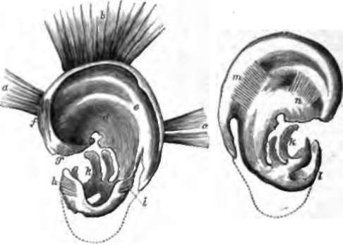
Fig. 120. Cartilage and Muscles of External Ear. A, Outer aspect. B, Cranial aspect, a, b, c, attrahens, attollens, and retra-hens aunculam muscles; d, concha; e, antihelix; f, g, large and small muscle of helix; h, tragus and tragic muscle; i, antitragus and antitragic muscle; k, the edge of the cartilage which is attached by fibrous tissue to the external auditory meatus of the temporal bone; l, tragus from behind; m, transverse muscle crossing the sulcus at the back of the antihelix; n, oblique muscle crossing sulcus at the back of the inferior branch of the antihelix. The lobule is represented in dotted outline.
Continue to: