187. Lachrymal Apparatus
Description
This section is from the book "Animal Physiology: The Structure And Functions Of The Human Body", by John Cleland. Also available from Amazon: Animal Physiology, the Structure and Functions of the Human Body.
187. Lachrymal Apparatus
The secretion of tears is primarily useful for keeping the surface of the cornea clear, and, in connection with this object, there are channels provided by which they may be removed without unduly accumulating or rolling over the cheeks. But the flow of tears is likewise influenced by the emotions; and any one who is familiar with the important effects which may be wrought by the action of one or two leeches, will be slow to doubt that a copious flood of tears may be of great utility in relieving a congested condition of the brain.
The lachrymal gland is similar in structure to the salivary glands, is about the size of an almond, is situated in the upper and outer angle of the orbit, under cover of the upper eyelid, and pours out its secretion by several ducts.
The eyelids are closely associated with the lachrymal gland in function. The upper lid is the more important of the two; it is larger than the lower, has twice as many eyelashes, has a special muscle for raising it, and is stiffened by a cres-centic plate of thin cartilage (superior tarsal cartilage) beneath its mucous membrane, while the lower lid has only a linear strip of that substance inside its margin. The upper and lower tarsal cartilages are united at the inner corner or canthus of the eye by a little tendon (tendo oculi) to the bone; and to the sides of this tendon are attached the fibres of the orbicularis palpebrarum muscle, a thin sheet of subcutaneous fibres which pass in circles round the eyelids, spreading over the adjacent parts of the cheek and forehead. This is the muscle by which the eyelids are closed; and its attachment to the tendo oculi explains why it is that, when the lids are forcibly shut, as, for example, by a reflex action on tasting something sour, their edges are drawn inwards to the nose. When the eyes are opened, the lower lid falls back into its place by elasticity, but the upper lid is raised by its levator palpebrae muscle, which comes forward from the back of the orbit, lying on the superior rectus, and is attached to the upper border of the superior tarsal cartilage. On everting either eyelid, a set of nodulated yellow streaks may be seen beneath the mucous membrane or conjunctiva (p. 229). They are the Meibomian follicles, and are a set of sebaceous glands which keep the margins of the lids oiled, and so help to prevent the tears from running over the cheeks.
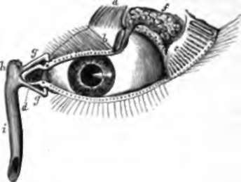
Fig. 125. Lachrymal Apparatus, a. Levator palpebae muscle; b, tarsal cartilago of the upper eyelid; c, Meibomian follicles, exhibited by division of the upper eyelid and reflection of the outer part; by, caruncula; c, plica semilunaris; f, lachrymal gland with the orifices of its ducts below it; g, canaliculi, with the puncta lachrymalia on the edges of the eyelids; h, lachrymal sac; i, nasal duct.
At the inner canthus of the eye, there is a little spongy-looking bit of mucous membrane, called the caruncula, resting on a fold of smoother membrane, the edge of which is laid against the eyeball. The fold is the plica semilunaris, and is the vestige or representative of a third eyelid, more distinct in many mammals than it is in man, and developed in birds, especially those of the nocturnal sort, as the membrana nictitans, a structure which rapidly sweeps across the eyeball to clear it, instead of the upper lid moving as it does when we wink. This can be readily seen in the owl.
Opposite the plica semilunaris, the margins of the eyelids, particularly that of the lower lid, change their direction; and on the elevation where this takes place there may be seen in each, when the lid is slightly pulled outwards, a little opening which rests against the eyeball. These openings are the puncta lachrymalia, which lead into two minute ducts called canaliculi, each of which passes vertically into the eyelid for a very short distance, then turns abruptly inwards to open into a sac behind the tendo oculi, whence the tears pass downwards by the nasal duct (p. 37) into the nose. Thus the tears, secreted by the lachrymal gland, pass across the eye, washing the conjunctiva; and every time a wink takes place, the puncta lachrymalia, and the parts of the canaliculi in connection with them, are lightly pressed against the eye, so that when the pressure is removed, the moisture is sucked into their interior; and only when there is a redundant secretion, or when by some irregular movement, as in laughter, the puncta lachrymalia are disarranged, do the tears accumulate within the lids, and overrun the cheeks.
Continue to: