150. Cranial Nerves (Fig 99)
Description
This section is from the book "Animal Physiology: The Structure And Functions Of The Human Body", by John Cleland. Also available from Amazon: Animal Physiology, the Structure and Functions of the Human Body.
150. Cranial Nerves (Fig 99)
From the under surface of the brain, a number of nerves emerge which are termed cranial. They differ greatly among themselves, both in size and function, and are variously numbered by different anatomists. But none of the methods of enumeration have the smallest title to be considered scientifically accurate; for they all agree in attempting to reduce to a linear series structures which do not serially correspond; therefore it is well to be guided by motives of convenience, and follow the plan generally adopted by English writers.
The first pair, the olfactory nerves, devoted to the sense of smell, are brushes of exceedingly soft and delicate filaments, given off from the under surface of the olfactory bulbs already alluded to as lying beneath the anterior lobes of* the cerebral hemispheres. These olfactory bulbs are much more largely developed in the majority of mammals than in man, and in some of them have cavities communicating with the interior of the brain. They are, in fact, vesicular outgrowths from the brain.
The second pair, the optic nerves or nerves of sight, come off, as has been already explained, from the optic commissure. Development shows that they also are vesicular outgrowths from the brain.
The third, fourth, and sixth pairs are all of them small motor nerves, which supply muscles of the eyeball, the third and fourth appearing above the pons Varolii, and the sixth below it.
The fifth pair, the trifacial nerves, are large trunks, whose fibres take origin in the medulla oblongata, and pierce the pons Varolii. They resemble the spinal nerves, in consisting each of a motor and sensory root; and in the sensory, which is much the larger, having a ganglion on it. The sensory part separates into three divisions, and supplies the scalp in front of the ear, and all the face, as well as the teeth and a large part of the tongue, with sensation; the motor part mixes with the third division of the sensory, and is distributed to the muscles of mastication.
The seventh pair consists of two portions, which emerge on the surface of the brain, in the angle between the crus cerebri, the cerebellum, and the medulla oblongata, and enter the temporal bone together. One portion, the portio mollis or auditory nerve, is the nerve of hearing, and is distributed within the bone; the other, the portio dura or facial nerve, is conducted through the temporal bone, and proceeds to the face to supply the muscles of expression. Within the temporal bone, the portio dura gives off the chorda tympani, which traverses the tympanic cavity, and joining with the lingual branch of the fifth nerve passes to a small ganglion, the submaxillary ganglion, and is the nerve alluded to at p. 64 as governing the secretion of the submaxillary gland.

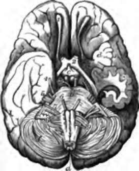
Fig. 99. Under Surface of Brain. a, Spinal cord cut across below the medulla oblongata: on the latter are seen, from the middle line outwards, anterior pyramids, olivary bodies, and restiform bodies. b, Pons Varolii; c, infundibulum (the pituitary body having been removed); d, gyri operti, or island of Reil; e, section of descending cornu of the left lateral ventricle, displayed by removal of part of the middle lobe of the hemisphere, exhibiting sections of the tŠnia hippocampi and hippocampus major; f, cerebellum. 1, Olfactory bulb; 2-9, successive cranial nerves, marked each with its proper number.
The eighth pair comprises three pairs of trunks, which arise from the medulla oblongata, and leave the skull by one pair of apertures. (1.) The glossopharyngeal nerves supply sensory branches, devoted to the sense of taste, to the back of the tongue, and motor and sensory branches to the pharynx. (2.) The pneumogastric or vagus nerve is both sensory and motor; it descends on the aesophagus to the stomach, and filaments may be traced from it even to the viscera. It supplies, in its course, branches to the pharynx, larynx and lungs, and the heart, and is intimately associated with the sympathetic, in connection with which it will be again referred to. (3.) The spinal accessory is entirely motor, and. consists of two parts, of which one, the accessory, joins the pneumogastric, while the other is distributed to two large muscles, the sterno-mastoid and trapezius.
The ninth pair, or hypoglossal nerves, are the motor nerves of the tongue. They emerge from the medulla oblongata behind the olivary eminences, which separate them from the eighth pair in front of these eminences.
Continue to:
- prev: The Whole Of The Brain Above The Level Of The Tentorium. Continued
- Table of Contents
- next: 151. Functions Of The Encephalon
