Spine And Acromion
Description
This section is from the book "The Anatomy Of The Human Skeleton", by J. Ernest Frazer. Also available from Amazon: The anatomy of the human skeleton.
Spine And Acromion
This has a thick rounded bar at its free outer border, which is the only part of the process to be preformed in cartilage (Fig. 58). The remaining thinner portion is apparently ossified directly in membrane, which represents probably the distal attachment of part of the Trapezius.
The upper and lower borders are continued behind into the aponeurotic rims for the spinatus muscles on the vertebral edge : this indicates that the covering aponeuroses are attached to the spine. Examine the areas for Trapezius and Deltoid : in both cases they spread on to the superficial surface of the bone for some distance, but in the case of the Deltoid the area narrows to the line of the border in its inner half, and so corresponds with the fact that the most posterior fibres of the Deltoid are aponeurotic, lying on and fused with the infraspinous aponeurosis, and extending on this nearly to the vertebral border of the bone. The Trapezius area extends in along the spine till it lies above the deltoid tubercle, where it narrows and turns downwards and outwards, forming a hook-shaped roughened ridge. This arrangement of the insertion-which is really responsible for the existence of the " deltoid tubercle "- is due to the'convergence of the fibres of Trapezius (Fig. 59), which would otherwise be twisted and overlap one another as they reached their insertion. The inner part of the spine is covered by Trapezius, which plays over it, and a small bursa may be found here very occasionally. The angle of the acromion overhangs the Infraspinatus, which plays against its deep surface. In some animals, as in the rabbit, the angle is drawn out into a long " metacromial " process.
Observe that the facet for the clavicle occupies the greater part of the inner border of acromion, so that Trapezius only reaches a very little part of this border before passing on to the clavicle, whereas the Deltoid arises along the whole of the outer border and then along the front before it reaches the clavicle. Also observe that both muscles have comparatively broad markings for their attachments, which are evidently by mixed muscle and tendon, and moreover the Deltoid area along the outer border has four ridges on it which indicate the places of attachment of the intramuscular tendons in this muscle (Figs. 57 and 59).
The facet is obliquely cut, facing upwards and inwards, so that the clavicle is placed on a slightly higher level and tends to ride over the acromion : this explains the difficulty in retaining the bone in position when dislocation has occurred between them. The rough area outside the facet is for ligaments which are strengthened by fibres from Trapezius and Deltoid, thus being made stronger than the ligaments on the inferior aspect.
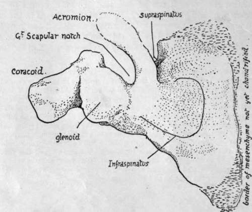
Fig. 58.-Dorsal surface of scapula in the sixth week.* From a model. There is no suprascapular notch because there is no supraspinous fossa. The cartilaginous acromio-spinous bar is distinct ; the interrupted line at its end indicates that it is lost in a condensation mass which will chondrify later to form the acromion. The glenoid cavity is partly on the coracoid. Note the small size of the areas for Infraspinatus and Supraspinatus.
The remaining upper surface of the process is subcutaneous, and has ramifying on it the small arteries of the " acromial rete." Its lower surface is smooth and is lined in part by the subacromial bursa which separates it from the tendon of Supraspinatus (Figs. 51 and 57, 5) : near the tip, in front, this surface has a slightly marked roughness for the attachment of the coraco-acromial ligament. The tendon and bursa separate it from the capsule of the joint, over which it is slightlv arched from before backwards.
The acromion is an epiphysis formed by centres occurring in the condensation in the middle of which the embryonic "spine" ends (Fig. 58). Sometimes the epiphysis does not join the spine by bone, and in such a case the cleaned bone shows a separated acromion: the l'ne of separation runs as a rule from in front of the angle to the bark of the clavicular facet, but may be much further forward, suggesting that the epiphysis has two centres at least for its formation, so that one or both of the pieces so developed may fail to join.
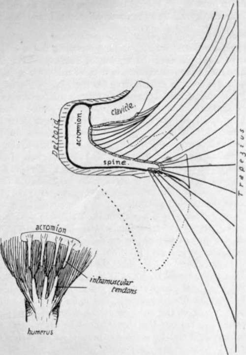
Fig. 59.-The larger drawing is a scheme to show how the disposition of the fibres of Trapezius accounts for the shape of its marking on the spine. The figure also shows the comparative attachments of this muscle and Deltoid on the scapula. The small figure is a scheme of the tendinous intersections or slips in the Deltoid at its origin and insertion, with the disposition of the fibres of the muscle ; this only affects the acromial part, on which most strain is thrown. The slips of origin make secondary markings on the acromion.
Glenoid Cavity
The rim affords attachment all round to the glenoid ligament (B), which is partly continuous above with the origin of the long head of Biceps from the bicipital tubercle : this is really on the base of the coracoid. The rim presents a notch in its upper and front part : this is better marked in the early state of the bone, indicating the junction of the " coracoid " and " scapular " parts of the articular surface, and the part above the notch has a separate centre of ossification. The oval cavity is very shallow, but in the recent state is deepened and enlarged by the glenoid ligament and by the fact that the cartilage hning it is thinnest centrally. The capsule is fastened to the outside of the rim and to the outside of the glenoid ligament, so that synovial membrane passes off this last on to the capsule with only a slight furrow intervening.
At the top, however, this arrangement is somewhat modified : the glenoid ligament not only joins the Biceps tendon, but is continued on the rim below it, and the capsule is attached in front of and behind the Biceps, passing up to become continuous with the coraco-humeral ligament on the coracoid process immediately above it.
Continue to: