Humerus. Part 4
Description
This section is from the book "The Anatomy Of The Human Skeleton", by J. Ernest Frazer. Also available from Amazon: The anatomy of the human skeleton.
Humerus. Part 4
Fig. 67.-The two planes of the Triceps. In "the first figure the inner head is seen as the deep plane of the muscle ; observe the musculo-spiral nerve crossing the inner head to reach the bone externally, a consequence of the height to which the origin of the head extends. In the next figure the other heads are seen lying on the deep plane, and a piece of the long head is cut away to show the musculo-spiral nerve lying at first between the long and inner heads before passing between the inner and outer. The ulnar nerve is applied to the inner head from the time it enters the arm, only separated from it by the musculo-spiral and superior profunda. The Deltoid is indicated by thick lines ; the circumflex nerve appears between long and outer heads deep to the muscle, so that each of its divisions must cross one of these heads under cover of the Deltoid.
The anterior circumflex artery runs outwards from the axillary sheath in the neighbourhood of the upper part of the I^atissimus dorsi tendon, thus reaching the floor of the bicipital groove on this tendon. It runs at first deep to the median and musculocutaneous nerves, then passes behind Coraco-brachialis and short head of Biceps, and in the groove is covered by the long head in its fibrous and serous coverings : it gives its bicipital or articular branch upwards in the groove, and some small foramina in the upper part of the groove receive minute twigs from this.
The artery itself proceeds transversely, crossing the outer lip above the pectoral tendon, and therefore piercing the upper expansion of the tendon : it now finds itself on the surface of the bone covered by the Deltoid.
Deltoid
The insertion is by strong tendon on the outer and front aspect of the shaft, but the posterior fibres tend to turn under the more external ones, and to reach the back and outer side of the bone at a slightly higher level : the roughness they cause on the bone may extend some distance up the back of the shaft. The front part of the muscle altogether covers the tendon of insertion of Pectoralis major (Fig- 65).
Triceps
Outer head: attached below to back of Deltoid tendon, and extending up to get just under cover of the lower border of Teres minor, a little distance internal to its insertion. If a thick hne is drawn from this point to the back of the deltoid impression it will mark the attachment, for the origin is by compressed tendon.
Inner head : extends upwards internally, passing under cover of Teres major for some I-f inch (Fig. 67). Sometimes these fibres are aponeurotic, making a mark on the bone just behind the Teres ridge, but they are frequently muscular and cause no roughness : the position of the muscle, however, is constant here. The area of origin is as shown in Fig. 63, and extends down farther on the outer than on the inner side. When the head is in position it is evident that the musculo-spiral nerve and superior profunda artery must lie on it and cross it to reach the groove, which is really outside this head of the muscle : this is the only part where these structures run in contact with the bone (see Fig. 67).
There are only two planes in the Triceps. The inner head, lying on and arising from the bone, is deep to the other two heads, which lie side by side behind it. Thus the musculo-spiral nerve, sinking back below the Teres major, disappears from view on the inner side of the arm by passing between the long and inner heads : it crosses the latter head, covered by the former, and then passes under cover of the outer head and reaches the bone outside the inner head. The circumflex nerve, passing back above the Teres major, goes between outer and long heads, and its anterior division crosses the former under cover of Deltoid while the posterior division crosses the long head, under the Deltoid to reach its lower border and turn round it. The ulnar nerve runs in a straight line from the Teres major along the inner side of the inner head just behind the internal intermuscular septum, and the septum reaches the prominent internal condyle, so the nerve passes behind the condyle, coming into contact with the bone here and passing on to the internal lateral ligament below.
External Intermuscular Septum is attached to the outer supracondylar ridge. If the ridge is traced up, it is found to lead toward the back of the deltoid impression, but is lost before reaching it owing to the depression of the spiral groove. The septum may be looked on as derived from the back of the Deltoid tendon, or, perhaps more truly, as a continuation down of the hne of origin of the outer head of Triceps from the point where it is attached to the back of the tendon. Brachiahs anticus arises from the bone in the angle between the hne of the septum and the Deltoid tendon, but is separated from the septum below this by the origin of Supinator longus (upper two thirds) and Extensor carpi radialis longior (lower one-third) from the front of the external ridge in front of the septum. The last-named muscle has a prominent marking on the bone.
Brachialis anticus shows individual variation to some degree in the distance to which its origin extends behind the Deltoid : also in its upward extension between Deltoid and Coraco brachialis, and if this is not great the musculocutaneous nerve may lie in contact with the bone for a short distance here before passing on to the muscle.
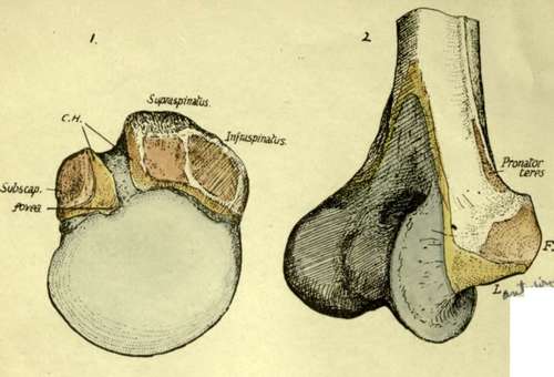
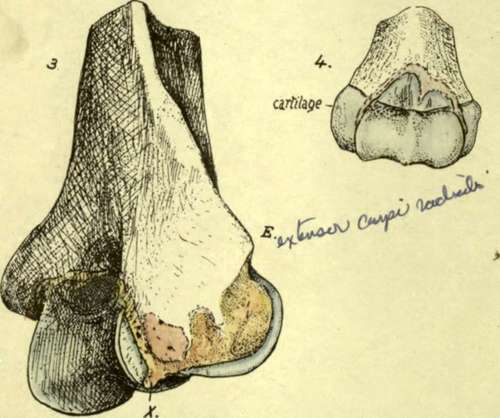
Fig. 68.-i. Upper end of right humerus from above. Shows the attachment of the coraco-humeral ligament, C.H., along the upper end of the bicipital groove or notch, and how it is continuous with the general capsule at the anatomical neck ; at this continuity in front the thin fibres of the superior gleno-humeral ligament, deep to the proper capsular fibres, reach the bone at the fovea humeri. 2. Front and inner view of lower end of right humerus. F. is the area of origin of the superficial flexors of the forearm. L. is the area of attachment of the anterior fasciculus of the internal lateral ligament ; this is continuous with the fibres of the anterior capsule at the edge of the trochlear eminence. The ligamentous structures do not encroach on the trochlea, which is therefore altogether covered by synovial membrane or cartilage ; the extent of the cartilaginous area is shown in Fig. 63. It can easily be appreciated that the anterior band of the lateral ligament is attached at L. in what is practically the axis of the movement of the ulna on the humerus, and hence this strong band can be tight in ordinary humero-ulnar movements and serve to maintain the bones in position. 3. The external condyle 01 the right humerus. The point of bone E. gives origin to the Extensor carpi radialis brevior and superficial Extensors of the forearm ; these also arise from the ligaments which are attached to the rough area immediately below E. The ligaments are shown in sitd in Fig. 72. The whole rough area is for the ligamentous tissue. The origin of Anconeus is generally larger than is shown in the figure. A", marks the attachment of the cruciate ligament. 4. Lower end of left humerus at birth, from the front. The whole of the lower end is cartilaginous. The capsule is attached partly on bone and partly on cartilage, so that separation of the cartilaginous end would involve opening of the joint. Observe the definite indication of the two parts of the composite joint cavity, radial and ulnar.
At its lower part, moreover, the area of origin of the muscle seems to vary in extent, but it always ends some little distance above the capsule. There is a large origin from the internal septum, but there can be none from the outer septum, except at its upper end.
The capsule of the elbow-joint makes a distinct marking on the bone, well above the coronoid and capitular fossa? in front, but across the floor of the olecranon fossa behind. The upper part of the olecranon fossa contains loose fatty tissue which is continuous, through deficiencies in the capsule leading to the subsynovial plane, with intra-articular fatty pads.
Continue to:
- prev: Humerus. Part 3
- Table of Contents
- next: Condyles