Phalanges Of The Foot
Description
This section is from the book "The Anatomy Of The Human Skeleton", by J. Ernest Frazer. Also available from Amazon: The anatomy of the human skeleton.
Phalanges Of The Foot
Compared with those in the hand, the phalanges of the foot in general are recognised at once (Fig. 85) by their thin and rounded shafts and large extremities : in the big toe they are broad and strong, but short for their size, and the first shows an obliquity of its base (Fig. 138), which enables one to place it at once on its proper side.

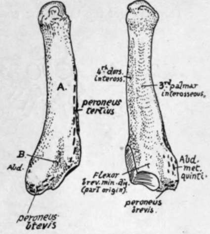
Fig. 156.-Left fifth metatarsal. Dorsa and plantar aspects. A. is the subcutaneous surface outside the dorsal ridge for Peroneus tertius ; a tendinous slip from Peroneus brevis passes on to this surface along the groove B., covered by ligamentous fibres attached to the margins of the groove.
The oblique direction of the first phalanx in the great toe does not appear to be the result of wearing boots : possibly it may be correlated with the outward turn of the foot as an adaptation to such acts as running or even walking, in which the body is lifted forward on the ball of the great toe, and such a direction of the phalanx keeps it out of the way and avoids over-extension of the joint. This does not, however, seem a very satisfactory explanation of the condition.
The account already given of the phalanges of the hand can be applied, mutatis mutandis, to those of the foot, but a few points must be noticed first. The middle phalanges get quickly smaller from within outwards, the fifth being usually an irregular nodule of bone ; the same in a less marked degree may be said of the last phalanges, and there is frequently a fusion between the middle and last bones in the little toe, and even occasionally in the other toes.
The bones of the tarsus are composed of cancellous tissue enclosed in very thin shells of compact bone. The cancellous lamellae are mainly directed in the lines of pressure transmitted from the tibia through the astragalus to the other bones : the arrangement is shown schematically in Fig. 157.
The correspondence of the foot with the generalised type of carpus and tarsus (p. 116) is seen in Fig. 157. In the same figure the hand and foot are compared to show the different components that " correspond " with each other.
Continue to:
