Ossification Of Femur
Description
This section is from the book "The Anatomy Of The Human Skeleton", by J. Ernest Frazer. Also available from Amazon: The anatomy of the human skeleton.
Ossification Of Femur
The bone is represented by a short and thick cartilaginous rudiment, in which ossification commences, at the centre of the future shaft, towards the end of the second month or earlier.
At birth the shaft is bony and the neck is short, while the trochanters and the extremities are in cartilage ; in the lower end, however, there is usually a small ossific centre in the depth of the cartilage, but this may not appear till just after birth.
The centre for the head appears in the first year, that for the great trochanter at three, and for the small trochanter at twelve or thirteen. Fusion of the head epiphysis with the neck, which has become longer, occurs at about eighteen, and the trochanteric epiphyses join the shaft about the same time or a little earlier. The bony lower end remains distinct until twenty-three or twenty-four, when its epiphysial line ossifies ; so the lower is the growing end of the bone.
The occasional centre for the gluteal ridge has already been mentioned (P- 147)-
The rough linea aspera becomes apparent at out puberty.
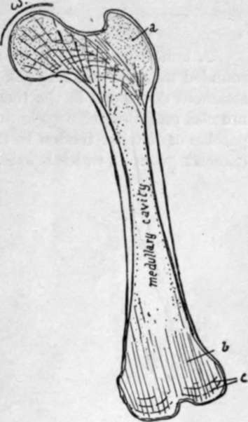
Fig. 125.-Scheme to illustrate the structure of the femur. The shaft is a hollow bony cylinder in the middle, but has cancellous tissue at the ends ; this is dense in the trochanter, a, but does not show any particular arrangement here, but this is not the case in the head, neck, and lower end, in the lines of weight-transmission. In the lower end there is a tendency to formation of vertical lamellae, 6, with cross-lamella; near the surfaces, c. In the neck there are spiral lamella; in the plane of the shaft wall, giving an appearance of arcades on section, and continued into dense cancellous bone in the head. Observe that the weight, m, will be transmitted from the upper part of the head through these spirals- to the lower wall of the neck ; here the wall is accordingly thick and noticeable on section, and is termed the calcar femorale.
The time of appearance of the centre for the lower end seems to be somewhat variable : as a rule it appears shortly before birth, but in a small number of cases it may be found as early as the eighth month, and in others, very rarely, it may be delayed even as long as the second year after birth.
Continue to:
- prev: Lower End
- Table of Contents
- next: Patella