The Renal Veins
Description
This section is from the book "Anatomy Of The Arteries Of The Human Body", by John Hatch Power. Also available from Amazon: Anatomy of the Arteries of the Human Body, with the Descriptive Anatomy of the Heart.
The Renal Veins
The Renal Veins, called also the emulgent veins, are two large vessels which escape, one from the hilus of each kidney. The right renal vein is shorter than that of the left, in consequence of the vena cava lying close to the right kidney; and it runs in a more oblique direction than the vein of the left side, because the right kidney is situated lower down than the left. The left renal vein is longer and takes a more transverse course than the right; it crosses in front of the aorta and the spine in order to reach the left side of the inferior vena cava; and in this part of its course it lies behind the third portion of the duodenum. Sometimes instead of passing in front of the abdominal aorta, it passes behind it: this vein receives the contents of the spermatic vein of the left testicle.
The branches given off by the renal arteries are, first, inferior capsular branches to the supra-renal capsules; secondly, branches to the surrounding areolar tissue and adipose membrane; thirdly, a small branch or two to the ureter, and, lastly, terminating branches. The terminating branches are disposed of in the manner which we shall now describe. Arrangement of the vessels in the kidney.Within the hilus or notch in the kidney the terminating branches first pass between the calyces, and then run in straight lines between the cones of the tubular structure till they arrive at the cortical structure of the organ. In the tubular portion, according to Mr. Toynbee, the minute arteries " are arranged in bundles in the shape of elongated cones whose bases are continuous with the cortical portion, and their apices directed towards the mammillary processes."* When these minute arteries have entered the cortical structure of the kidney, most of them terminate in forming small tufts of capillary vessels in the Malpighian corpuscles. These bodies have been described by Mr. Bowman as being formed in the first instance by a capsule which consists of an expansion of the dilated commencement of a urinary tubule; through this capsule a minute artery passes, called vas infer ens, which, after arriving within it, subdivides into several minute capillary branches, which form a number of vascular loops closely bound up together within the capsule, so as to form a tuft: from this tuft of vessels a small vein takes its origin, vas efferens, which escapes from the inner portion of the corpuscle, passes through the capsule and joins a plexus of veins, formed of several efferent veins, which are situated between the Malpighian corpuscles, and surround the small convoluted tubuli uriniferi immediately after their origin : these capillary veins terminate in the formation of the renal vein. Mr. Bowman describes a number of minute capillary arteries which do not go to the Malpighian corpuscles, but which envelope the convoluted tubes and communicate directly with the veins. According to Mr. Bowman, the capsule of the Malpighian body consists of the dilated origin of the urinife-rous tubule; whilst, according to Mr. Toynbee, this capsule consists of a structure totally distinct from the tubule, and which surrounds the convoluted origin of the tubule in conjunction with the arterial tuft and the commencement of the efferent vein.
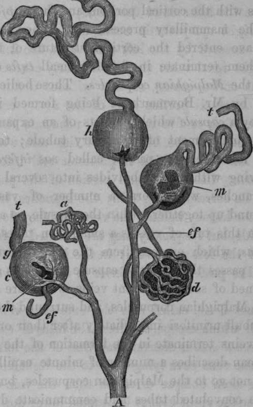
Fig. 40. Microscopical Anatomy of Kidney. Represents the Arrangement of the Vessels in the Malpighian Tuftsafter Bowman.
A, Arterial Branch, with its subrlivirions. At a, the Capsule is ruptured, and only some of the Vessels are seen. At d, the vessels are well filled, and the injection passed out through the efferent vessel ef. Ate, h, the injection has extravasated, and passed along the tube, ef, Efferent Vessel, m, m, The injection, on escaping into the capsule, has not spread over the whole tuft, t, t, t, Uri-niferous Tubes.
* Med. Ch. Trans., vol. xxix., 1846. Phil. Trans., 1842.
In the early period of intra-uterine life, the kidney is formed of a number of independent lobules, each supplied with a distinct set of vessels; and even in the adult there remains so much distinction, that different compartments of the kidney can be injected of different colors.
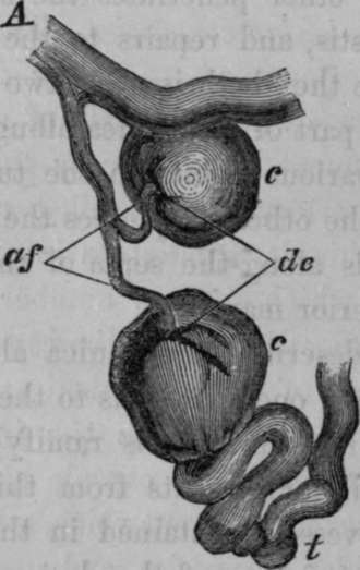
Fig. 41. Represents two Malpighian Bodies injected. The tufts are burst, and the fluid has escaped into the capsule. In one case it has passed also along the tube, the tortuosity of which, at its commencement, is well seen.-After Bowman.
A, Arterial Branch, a, f, Arterial Twigs or Afferent Vessels of the Malpighian Tufts, c, c, Malpighian Bodies, distended, d, e, The Depression sometimes seen where the Afferent and Efferent Vessels pass, t, Uriniferous tube.
Continue to: