III. Afferent Or Sensory Paths Of The Hunger Complex And The Question Of The Cerebral "Hunger Center"
Description
This section is from the book "The Control Of Hunger In Health And Disease", by Anton Julius Carlson. Also available from Amazon: The Control of Hunger in Health and Disease.
III. Afferent Or Sensory Paths Of The Hunger Complex And The Question Of The Cerebral "Hunger Center"
1. Role Of The Vagi
The vagi nerves are the main, if not the only, afferent pathway for the gastric hunger impulses, although Luciani assumes that some of the hunger impulses from the stomach are carried by the sympathetic (splanchnic) fibers. If the contraction of the small intestine contributes to the hunger sensation, the afferent hunger impulses may involve sympathetic and spinal nerves, but all sensory conduction from the stomach appears to be confined to the vagi, because no central reflexes of any kind can be evoked by stimulation of the stomach after section of all the vagi fibers to that organ (Miller). We need not here refer to the physiologists who have argued against the vagi nerves (and in favor of the splanchnics) as being concerned in hunger on the basis that animals will continue to eat after section of these nerves or after excision of the stomach, as these objections have already been considered and refuted.
2. The Primary Hunger Center
2. The primary hunger center is therefore the sensory nuclei of the vagi nerves in the medulla (fasciculus solitarius). Some of the more direct hunger reflexes (such as salivation, vasomotor fluctuations, etc.) may be carried out via these medullary centers alone. Luciani assumes also spinal hunger centers analogous to these sensory vagi nuclei. There is no evidence that the processes of conscious hunger sensation can take place in the medullary nuclei.
3. Role Of The Optic Thalami And The Mid-Brain
The important facts in this connection are the hunger behavior of decerebrated animals (acephalic infants, dogs, pigeons, frogs). This hunger behavior has already been ascribed. These animals minus the cerebral hemisphere, but with the thalamic region of the brain intact, exhibit practically all the hunger behavior of normal animals, except the intelligent search for and ingestion of the food. But even this statement requires limitation, for, according to Ewald,1 decerebrated pigeons and frogs will finally eat spontaneously if kept in good condition for a sufficient time (months) after the operation.
Rogers has recently made the important observation that the hunger behavior of the decerebrated pigeon is completely abolished on removal of the optic thalami. It is thus clear that this region contains important nuclei for the elaboration of the bodily responses to the hunger impulses from the stomach. Whether or not the processes of conscious hunger sensations are elaborated in the thalamus cannot be determined on experimental animals. L. R. Miiller assumes that conscious hunger sensations are evoked in the mid-brain. We have seen that hunger is essentially pain, and some neurologists take the position that the sensation of pain is a thalamic rather than a cortical function. This view is supported by the extensive studies of Head and Holmes on the change in the pain sense in persons with thalamic lesions and intact cortex. The frequent occurrence of excessive hunger or polyphagia in persons with tumors of the pineal glands have by some (Schuller) been interpreted as due to a pressure stimulation of subcortical hunger centers. Whether the thalamic processes caused by the gastric hunger contractions are conscious or merely subconscious reflexes, or whether the nuclei concerned with these processes are identical with those involved in pain sensation, it is clear that the thalamus is a very important reflex and relay station for the afferent hunger impulses.
1 Quoted from A. L. Gillespie, The Natural History of Digestion, New York, 1898,
4. Cortical Hunger Centers
Concerning these practically nothing is known. There can be little doubt that conscious hunger involves in some way cortical processes, and one might expect the part of the cortex involved would be contiguous with that for the gustatory sense, the latter being placed in the hipochampal gyrus by the majority of neurologists and psychiatrists. Roux assumes that the cortical hunger center is in the Rolandic area, thus supporting Bechterew, who locates the conscious taste processes in the region of the Rolandic area which innervates the muscles of mastication and deglutition.
To recapitulate: In the afferent phase of the hunger complex the facts clearly established are the rdle of the vagi, and the sensory vagi nuclei in the medulla, and the great importance of the thalamus. The cortical factors in hunger are unknown, and the same applies to the detailed roles of the subcortical hunger centers both in health and in disease. This field of the physiology of hunger is therefore mainly "gaps and guesses." It remains for the clinical investigator to correct the guesses and fill up the gaps, as very little can be done with these problems on animals below man, at least with the methods so far available to the physiologist.
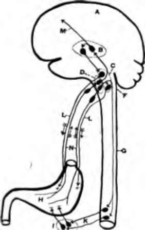
Fig. 26.-Diagram of the nervous mechanism of the hunger sense. A, cerebrum. B, optic thalami. C, motor nuclei of the vagi nerves. D, sensory nuclei of the vagi nerves. F, medulla oblongata. G, spinal cord. H, stomach. /, visceral sympathetic ganglia. K, splanchnic nerves. L, motor fibers to the stomach, if, sensory paths (hunger) to cerebrum (hypothetical). N, sensory fibers from stomach in the vagi. + indicates motor, - indicates inhibitory effects, -> indicates direction of nervous conduction.
Continue to:
- prev: 3. Results On Man
- Table of Contents
- next: Chapter XIII. The Chemical Control Of The Hunger Mechanism. I. Analysis Of The Problem