Minute Anatomy Of Hevea Bark
Description
This section is from the book "Rubber And Rubber Planting", by R. H. Lock. Also available from Amazon: Rubber And Rubber Planting.
Minute Anatomy Of Hevea Bark
The networks of laticiferous vessels in Hevea are seen in transverse section to be separated in the radial direction by about five rows of cells and other elements. Many of these are actually the sieve tubes and companion cells of the phloem. No radial connections have been observed between the several networks, which appear to extend completely round the stem, being thus concentric and having no communication with one another. The arrangement of the cells composing the networks is seen more clearly in longitudinal sections. In the younger networks, close to the cambium, the remains of the cell-walls may still be seen partly blocking the interior of the vessels; in the older vessels the transverse and most of the longitudinal walls between adjoining cells become absorbed. Two stages in the dissolution of the cell-walls are seen in Figs. 8 and 9. Fig. 8 represents a tangential section of a network very close to the cambium, whilst that shown in Fig. 9 is older and further away.
In the seedling the nodes of the network may be partly formed by tubular outgrowths of the cells which meet and fuse together, thus recalling the behaviour of latex tubes proper. Fig. 10 shows such a fusion in a transverse section of the seedling. But in the secondary laticiferous tissue derived from the cambium there is hardly any outgrowth of this kind. The complete network is laid down in situ.
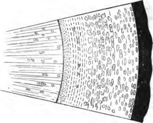
Fig. 7. Hevea transverse section of bark x 5. The tangential lines represent groups of latex vessels.
In material which has been preserved in strong Flemmings solution, the contents of the cells which are about to form the latex vessels are distinguishable almost immediately after they have been cut off from the cambium (Fig. 11). Their appearance and arrangement at this stage is closely comparable with that of the companion cells of the phloem. '"Sometimes the nuclei of the surrounding larger cells are to be observed closely pressed against the walls next to the incipient latex vessels, suggesting that the former may play some part in providing materials for the contents of the latter. The adult bark of Manihot has not been so closely studied as that of Hevea, but the manner of formation of the vessels appears to be closely similar in the two cases. For further details, and for a full account of the structure of the laticiferous system in the seedling, the reader is referred to the papers of Dr D. H. Scott.
Plate IV
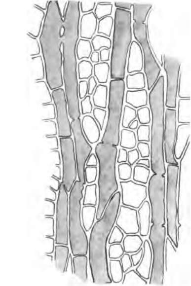
Fig. 8.
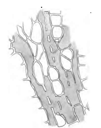
Fig. 9.
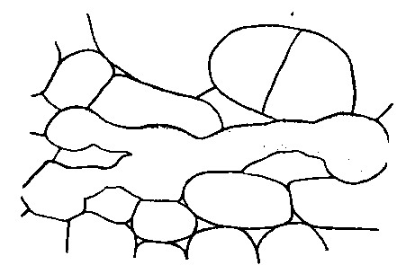
Fig. 10.
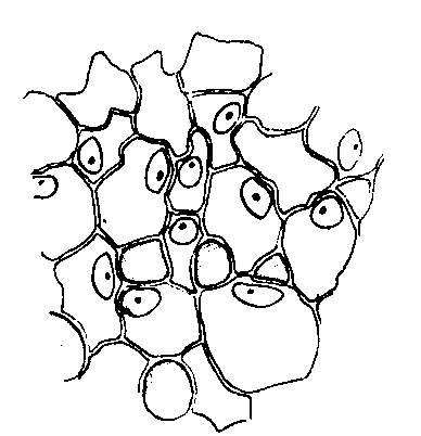
Fig. 11.
Anatomy of Hevea
Continue to: