The Inferior Radio-Ulnar Joint
Description
This section is from the book "Surgical Anatomy", by John A. C. MacEwen. Also available from Amazon: Surgical Anatomy.
The Inferior Radio-Ulnar Joint
The Inferior Radio-Ulnar Joint Consists of (a) an articulation between the head of the ulna and the sigmoid facet on the lower end of the radius, and (b) the articulation between the head of the ulna and the triangular fibro-cartilage of the wrist. The fìbro-cartilage is attached by its base to the inferior border of the radius, and by its apex to the depression at the base of the styloid of the ulna. It binds the bones together, and separates the inferior radio-ulnar joint from the wrist. The inferior radio-ulnar joint presents weak anterior and posterior ligaments, which extend from either side of the radial sigmoid notch to the ulna and triangular fibro-cartilage. The synovial membrane is loose, and lines both the articulation between radius and ulna and the interspace between the lower end of the ulna and the triangular fibro-cartilage. Pronation and supination movements take place round an axis from the head of the radius through the end of the ulna, to the fourth metacarpal. Pronation is limited by the lower two-thirds of the interosseous membrane, part of the posterior ligament of the wrist, and apposition of the bones. Supination is limited by the lower third of the interosseous membrane, the internal lateral ligament of the wrist, and contact of the posterior edge of the sigmoid cavity of the radius with the tendon of the extensor carpi ulnaris. Supination is the more powerful movement of the two.
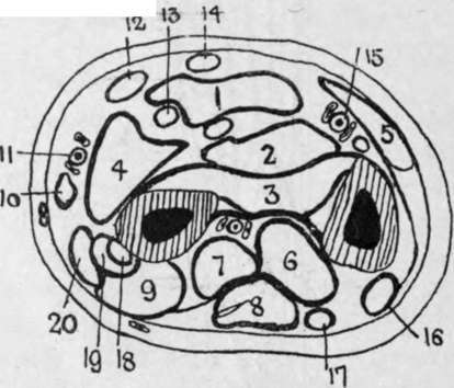
Fig. 55.-Outline Diagram of Transverse Section of Forearm in Lower Third.
(After Braune.)
1. | Flex, sublimis | 8. | Ext. comm. dig. | 15. | Ulnar vessels and nerve. |
2. | Flex, profund. | 9. | Ext. brev. poll. | 16. | Ext. carpi ulnaris. |
3. | Pronat. quad. | 10. | Sup. longus. | 17. | Ext. min. dig. |
4. | Flex. long. poll. | 11. | Radial vessels. | 18. | Ext. carpi rad. brev. |
5. | Flex, carpi ulnar. Ext. indicis. | 12. | Flex, carpi rad. | 19. | Ext. carpi rad. long. |
6. | 13. | Median nerve. | 20. | Abductor pollicis. | |
7. | Ext. long. poll. | 14. | Palmaris long. |
Continue to: