The Vessels And Nerves Of The Lower Extremity
Description
This section is from the book "Landmarks And Surface Markings Of The Human Body", by Louis Bathe Rawling. Also available from Amazon: Landmarks and Surface Markings of the Human Body.
The Vessels And Nerves Of The Lower Extremity
The gluteal artery emerges from the great sacro-sciatic notch, (Fig. XXI, 10.) above the pyriformis muscle, at the junction of the inner and middle thirds of a line drawn from the posterior superior iliac spine to the top of the great trochanter of the femur of the same side. The sciatic artery may be ligatured at a point which lies just external to the junction of the middle and lower thirds of a line drawn from the posterior superior iliac spine to the outer part of the ischial tuberosity of the same side. (Fig. XXI, 8.) This line also cuts across the posterior inferior iliac spine and the tip of the ischial spine, whilst the internal pubic artery lies immediately internal to the seat of election for ligation of the sciatic-artery.

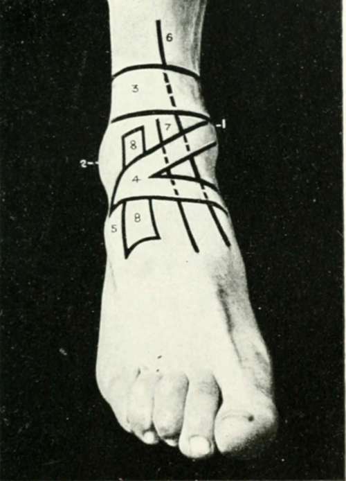
Fig. XXVI. The Region Of The Ankle And Foot
1. The internal malleolus.
2. The external malleolus.
3. The transverse band of the anterior annular ligament.
4. The Y-shaped hand of the anterior annular ligament.
5. The head of theos caicisand the extensor brevis digitortim.
6. The tibialis anticus synovial sheath.
7. The extensor longus hallucis synovial sheath.
8. The extensor longus digitorum and peroneus tertius synovial sheath.

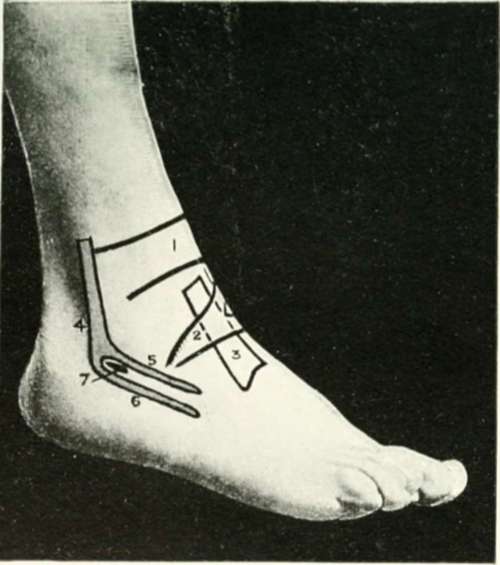
Fig. XXVIII. The Region Of The Ankle And Foot
1. The transverse limb.
2. The stem of the Y-shaped part of the anterior annular ligament.
3. The extensor longns digitorum and peroneus tertins synovial sheath.
4. The peroneus longus and brevis synovial sheath.
5. The peroneus brevis sheath.
6. The peroneus longus sheath.

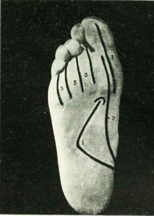
Fig. XXIX. The Region Of The Ankle And Foot
1. The external plantar artery.
2. The internal plantar artery.
3. 3. The distal flexor synovial sheaths
Continue to:
- prev: The Anterior Annular Ligament Of The Ankle
- Table of Contents
- next: The Common And Superficial Femoral Arteries
