The Pancreas
Description
This section is from the book "Landmarks And Surface Markings Of The Human Body", by Louis Bathe Rawling. Also available from Amazon: Landmarks and Surface Markings of the Human Body.
The Pancreas
The head of the pancreas occupies the concavity of the duodenal loop, (Fig. XIX, 4.)the body crossing the middle line at the level of the first and second lumbar vertebras, and occupying, therefore, the upper two-thirds of the space between the transpyloric (first lumbar) and subcostal (third lumbar) planes. The tail of the pancreas extends to the left as far as the hilum of the spleen.
The small intestine is about 23 feet in length, the upper two-fifths being known as the jejunum, the lower three-fifths as the ileum.
The mesenteric attachment of the small gut extends from a point 1 to 1 1/2 inches to the left of the middle line, (Fig. XIX, 6.) and just below the transpyloric plane (duodeno-jejunal flexure), to the junction of the right lateral vertical and intertubercular planes. The line drawn between these two points should pass at first obliquely outwards and downwards to the right iliac fossa, curving finally outwards to the region of the ileo-caecal valve.
The ileo-caecal valve is placed opposite the junction of the right lateral vertical and intertubercular planes. (Fig. XIX, 7.)
The caecum is about 2 1/2 inches long, and the long axis of the sac is directed downwards, forwards, and inwards. (Fig. XIX, 8.)
This blind end of the large gut lies below the level of the intertubercular plane, and occupies the right iliac fossa and part of the right half of the hypogastric region.
X-ray pictures of the stomach filled with Barium show almost constantly a stomach of very different shape to that depicted in Fig. XIX. This figure has, therefore, been criticised. I have, however, had the opportunity of seeing many normal stomachs, both in the anatomical department and in surgery. The cardiac and pyloric orifices, under normal conditions, are more or less fixed points. The stomach itself varies in shape and position from hour to hour, according to the period of digestion. Some average, however, has to be taken, and I submit that the normal stomach is represented with sufficient accuracy.
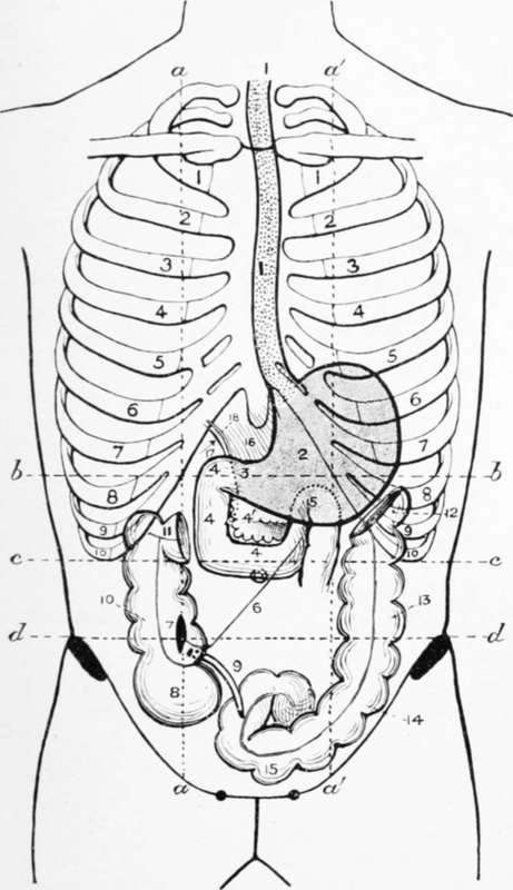
Fig. XIX. The Alimentary Canal
1. The oesophagus.
2. The stomach.
3. The pylorus.
4. The three parts of the duodenum and The pancreas.
5. The duodeno-jejunal flexure.
6. The attachment of the mesentery of the small intestine.
7. The ileo-caecal valve.
8. The caecum.
9. The vermiform appendix.
10. The ascending colon.
11. The hepatic flexure.
12. The splenic flexure.
13. The descending colon.
14. The iliac colon.
15. The ileo-pelvic colon.
16. The gastro-hepatic omentum.
17. The foramen of Winslow.
18. The common bile-duct.
N.B
The transverse colon has been omitted intentionally.
a, a, and a', a' = the lateral vertical planes.
b, b. The transpyloric plane.
c, c. The- subcostal plane.
d, d. The intertubercular plane.
Continue to: