Chapter V. The Lower Extremity
Description
This section is from the book "Landmarks And Surface Markings Of The Human Body", by Louis Bathe Rawling. Also available from Amazon: Landmarks and Surface Markings of the Human Body.
Chapter V. The Lower Extremity
The pubic spine, the iliac crest, the anterior and posterior superior iliac spines, and the iliac tubercles have all been previously alluded to and located. It has also been stated that a line uniting the two posterior superior iliac spines cuts across the spine of the second sacral vertebra. Below this line the remaining sacral spines and the coccyx are easily felt, (Fig. XXI, 4,5) though the coccyx itself is more readily verified by inserting the forefinger into the rectum whilst the thumb is placed over the bone externally.
The Region Of The Hip
(a) Posterior and external aspect. The great trochanter of the femur and the ischial tuberosity must now be examined. The latter process lies under cover of the lower part of the gluteus maximus muscle, (Fig. XXI, 7.) whilst the trochanter is subcutaneous in its lower part, and covered over in front and above by the insertion of the gluteus medius muscle.
The trochanter is more or less quadrilateral in shape, (Fig. XXI, 9.)fading off into the shaft of the femur below, (Fig. XXIII, 6.) and presenting a well-marked posterior border; the highest point of the trochanter corresponds to the posterior superior angle of the quadrilateral. When the body is in the erect position, there is a well-marked depression situated above and behind the great trochanter, which becomes less marked when, as the result of disease or disuse, the gluteus maximus undergoes atrophic changes. The fold of the nates does not correspond to the lower border of the gluteus maximus muscle, as it crosses almost transversely the lower oblique fibres of that muscle. This fold also becomes less distinct when the glutei muscles degenerate. The head and neck of the femur form with the shaft of that bone an angle of 125 to 130 degrees.
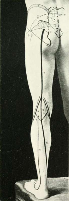
Fig. XXI. The Back Of The Thigh And Leg
1. The iliac crest.
2. The posterior superior iliac spine.
3. The sacrum.
4. The second sacral spine.
5. The coccyx.
6. The pyriformis muscle.
7. The ischial tuberosity.
8. The sciatic artery and the ischial spine.
9. The great trochanter of the femur.
10. The gluteal artery.
11. Nelaton's line.
12. The great sciatic nerve.
13. The internal popliteal nerve.
14. The external popliteal nerve.
15. The biceps tendon.
16. The semimembranosus muscle.
17. The semitendinosus muscle.
18. The outer head of the gastrocnemius muscle.
19. The inner head of the gastrocnemius muscle.
20. The posterior tibial artery and nerve.
21. The external saphenous vein.
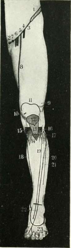
Fig. XXII. The Front Of The Thigh And Leg
1. The iliac crest.
2. The pubic spine.
3. Poupart's ligament.
4. The anterior crural nerve.
5. The common femoral artery.
6. The common femoral vein.
7. The crural canal.
8. The superficial femoral artery.
9. Vastus externus.
10. Vastus internus.
11. The upper limit of the synovial membrane of the knee-joint.
12. The patella.
13. The ligamentum patellae.
14. The tubercle of the tibia.
15. The internal tuberosity of the tibia.
16. The external tuberosity of the tibia.
17. The head of the fibula.
18. The internal saphenous vein.
19. The anterior tibial artery.
20. The anterior tibial nerve.
21. The musculo-cutaneous nerve.
22. The dorsalis pedis artery.
23. The dorsal venous arch.
Nelaton's Line
(Fig. XXI, 11., Fig. XXIII, 5.) "If in the normal state you examine the relation of the great trochanter to the other bony prominences of the pelvis, you will find that the top of the great trochanter corresponds to a line drawn from the anterior superior iliac spine of the ilium to the most prominent point of the tuberosity of the ischium. This line also runs through the centre of the acetabulum. The extent of displacement in dislocation or in fracture is marked by the projection of the trochanter behind and above this line " (Nelaton).
Continue to: