The Small Intestine
Description
This section is from the book "The Human Body: An Elementary Text-Book Of Anatomy, Physiology, And Hygiene", by H. Newell Martin. Also available from Amazon: The Human Body.
The Small Intestine
The Small Intestine commences at the pylorus and ends, after many windings, in the large. It is about twenty feet (six meters) long and about two inches (five centimeters) wide at its gastric end, narrowing to about two thirds of that width at its lower portion. Externally there are no lines of subdivision on the small intestine, but anatomists arbitrarily describe it as consisting of three parts, the first twelve inches being the duodenum, the succeeding two fifths of the remainder the jejunum, and the rest the ileum.
Why is it that an over-distended stomach sometimes causes palpitation of the heart?
Where does the small intestine commence? Where does it end? Describe its length and diameter. Of what divisions do anatomists describe it as consisting?
The Mucous Coat Of The Small Intestine
This is pink, soft, and extremely vascular. It is throughout a great portion of the length of the tube raised up into permanent transverse folds in the form of crescentic ridges, each fold running transversely for a greater or less way round the intestine (Fig. 50). These folds are the valvul conniventes. They are first found about two inches from the pylorus, and are most thickly set and largest in the upper half of the jejunum, in the lower half of which they become gradually less conspicuous; they finally disappear altogether about the middle of the ileum. The folds of the mucous membrane serve to greatly increase its surface both for absorption and secretion, and they also delay the food in its passage; it collects in the hollows between them, and so is longer exposed to the action of the digestive liquids.
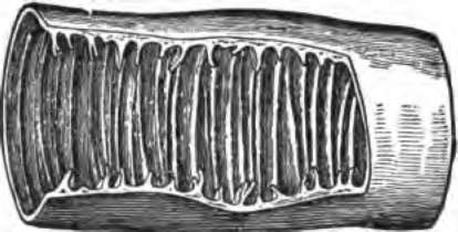
Fig. 50. A portion of the small intestine opened to show the valvul conni-ventes.
The Villi
Examined closely with the eye or, better, with a hand lens, the mucous membrane of the small intestine is seen not to be smooth but shaggy, being covered everywhere (both over the valvul conniventes and between them) with closely packed minute elevations standing up somewhat like the " pile" on velvet and known as the villi (Fig. 51). In structure a villus is somewhat complex. Covering it is a single layer of cells, beneath which the villus may be regarded as made up of a framework of connective tissue supporting the more essential constituents. Near the surface is a network of plain muscular tissue. In the centre is an offshoot of the lymphatic or absorbent system, sometimes in the form of a single vessel with a closed dilated end, and sometimes as a network formed by two main vessels with cross-branches. During digestion these lymphatics are filled with a milky white liquid absorbed from the intestines, and they are accordingly called the lacteals. They communicate with larger branches in the outer coats of the intestine, and these end in trunks which join the main lymphatic system. Finally, in each villus, outside its lacteals and beneath its muscular layer, is a close network of bloodvessels.
Give the general characteristics of the mucous membrane of the small intestine. What are the valvul conniventes? Where do they commence?. Where are they most developed? Where do they cease? What purposes do they subserve? What are the villi?
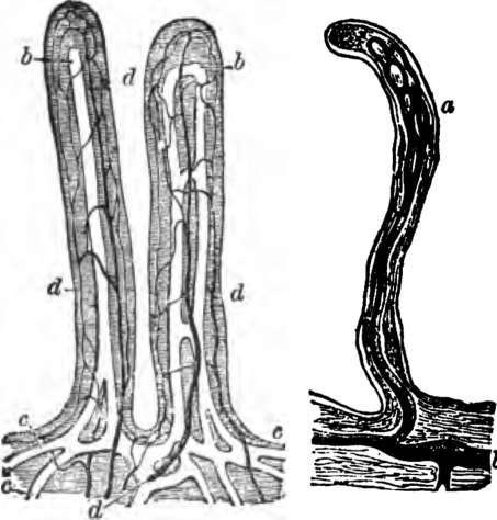
Fig. 51. Villi of the small intestine; magnified about 80 diameters. In the left-hand figure the lacteals. a, b, c, are filled with white injection: d, blood-vessels. In the right-hand figure the lacteals alone are represented, filled with a dark injection. The epithelium covering the villi, and their muscular fibres are omitted.
Describe the structure of a villus. What is found in its lymphatics during digestion?
The Glands Of The Small Intestine
Opening on the surface of the small intestine between the bases of the villi are small glands, the crypts of Lieberkühn. Each is a simple unbranched tube, lined by a single layer of cells.
The Muscular Coat Of The Small Intestine
The Muscular Coat Of The Small Intestine lying outside the mucous coat, is composed of plain muscular tissue, disposed in two layers: an inner circular, and an outer longitudinal. By their combined and alternating contractions they slowly force the digesting food along the tube.
In the duodenum are found in addition minute glands, the glands of Brunner, which lie outside the mucous membrane, and send their ducts through it to open on its inner surface.
Continue to: