The Microscopic Structure Of The Retina
Description
This section is from the book "The Human Body: An Elementary Text-Book Of Anatomy, Physiology, And Hygiene", by H. Newell Martin. Also available from Amazon: The Human Body.
The Microscopic Structure Of The Retina
The Microscopic Structure Of The Retina is very complex; although but 1/80 inch in thickness it presents ten distinct layers.
What is the form of the globe of the eye? Of what does it consist? Describe the outer coat. The second coat. What is the retina?
* In pink-eyed rabbits and in the pink-eyed ladies of " dime museums" this pigment is absent.
Beginning (Fig. 89) on its front or inner side we find, first, the internal limiting membrane, 1, a thin structureless layer. Next comes the nerve-fibre layer, 2, formed by radiating fibres of the optic nerve; third, the nerve-cell layer, 3; fourth, the inner molecular layer, 4, consisting partly of very fine nerve-fibrils, and largely of connective tissue; fifth, the inner granular layer, 5, composed of nucleated cells, with a small amount of protoplasm at each end, and a nucleolus. These granules, or at any rate the majority of them, have an inner process running to the inner molecular layer, and an outer running to, 6, the outer molecular layer, which is thinner than the inner. Then comes, seventh, the rod and cone fibre layer, 7, or outer granular layer, composed of thick and thin fibres on each of which is a conspicuous nucleus with a nucleolus. Next is the thin external limiting membrane, 8, perforated by apertures through which the rods and cones, 9, of the ninth layer join the fibres of the seventh. Outside of all, next the choroid, is the pigmentary layer, 10. The nerve-fibres are believed to be continuous with the rods and cones. Light entering the eye passes through the transparent retina until it reaches the rods and cones and excites these, and they stimulate the nerves.
The action of the light is probably in the first instance chemical. The rods are stained by a purple substance, which is bleached by light and regenerated in the dark. In the healthy eye the purple is reproduced nearly as fast as destroyed, by the pigment-cells of the retina. Parts of the retina which contain none of this vision purple can see,but they may possess uncolored substances which are changed by light.
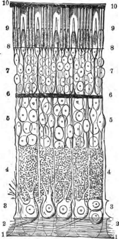
Fig. 89. A section through the retina from its anterior or inner surface, 1 in contact with the hyaloid membrane, to its outer, 10, in contact with the choroid. 1, internal limiting membrane; 2, nerve-fibre layer; 8, nerve-cell layer; 4, inner molecular layer; 5, inner granular layer; 6, outer molecular layer; 7, outer granular layer; 8, external limiting membrane; 9, rod and cone layer; 10, pigment-cell layer.
Describe the microscopic structure of the retina. On what constituents of the retina does light first act, in producing a sensation?
With what are the rods of the retina stained? How is the pigment reproduced?
Continue to: