The Kinds Of Glands
Description
This section is from the book "The Human Body: An Elementary Text-Book Of Anatomy, Physiology, And Hygiene", by H. Newell Martin. Also available from Amazon: The Human Body.
The Kinds Of Glands
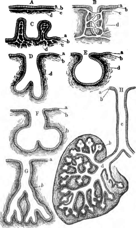
All portions of the body making and pouring forth secretions are not technically called glands. In the peritoneum, which lines the inside of the abdominal cavity (p. 11), we find simply a thin membrane (A, Fig. 40), having on its side nearer the cavity which it surrounds a layer of cells, a, and on its deeper side a network of very fine blood-vessels, c, supported by connective tissue, d. Such simple, smooth, secreting surfaces are not common; in most cases an extended area is required to form the necessary amount of secretion, and if this were attained simply by spreading out flat membranes, these, from their number and extent, would be hard to pack conveniently in the body. Accordingly, in most cases, a large area is obtained by folding the secreting surface in various ways so that a wide surface can be packed in a small bulk, just as a Chinese paper lantern when shut up occupies much less space than when extended, although the actual area of the paper in it remains of the same extent. In a few cases the folding takes the form of protrusions into the cavity of the secreting organ, as indicated at 0, Fig. 40, but much more commonly the surface extension is attained in another way, the supporting or basement membrane, covered by its epithelium, being pitted in or involuted as at B. Such a secreting organ is known as a true gland.
Why do we speak of different kinds of digestion? Illustrate. What is a gland? Give examples of glands. How do glands differ? What are the two chief types of glands named?
Name and describe a secreting surface which is not technically called a gland. What is gained by folding a secreting surface?

Fig. 40. Forms of Glands. A, a simple secreting surface ; a, its epithelium; 6, basement membrane ; c, capillaries ; B, a simple tubular gland ; C, a secreting surface increased by protrusions ; E. a simple racemose gland : D and G, compound tubular glands; F, a compound racemose gland. In all but A, B, and C the capillaries are omitted for the sake of clearness. H, half of a highly developed racemose gland ; c, its main duct.
Continue to:
