Diagram Of The Circulatory Organs
Description
This section is from the book "The Human Body: An Elementary Text-Book Of Anatomy, Physiology, And Hygiene", by H. Newell Martin. Also available from Amazon: The Human Body.
Diagram Of The Circulatory Organs
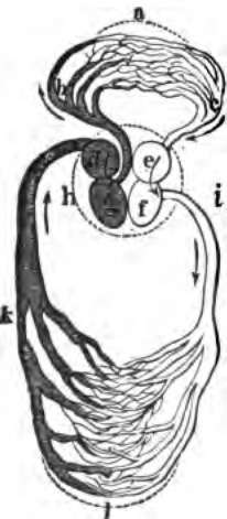
The general relationship of heart, arteries, capillaries, and veins may be gathered from the accompanying diagram (Fig. 57). The heart is essentially a bag with muscular walls, and internally divided into four chambers (d, g, e, f). Those at one end (d and e) receive blood from vessels opening into them and known as the veins. From there the blood passes on to the remaining chambers (g and f), which have very muscular walls and, forcibly contracting,drive the blood out into vessels (i and b) which communicate with them and are known as the arteries. The big arteries divide into smaller; these into smaller again (Plate IV), until the branches become too small to be traced by the unaided eye, and these smallest branches end in the capillaries, through which the blood flows and enters the commencements of the veins; the veins convey it again to the heart. At certain points in the course of the blood-path valves are placed, which prevent a back-flow. This alternating reception of blood at one end of the heart and its ejection from the other occurs about seventy times a minute during health.
By comparison with the water pipes of a house. What is the use of heart, arteries, and veins? When does the blood do its physiological work?

Fig. 57. The heart and blood-vessels diagrammatically represented.
Sketch on the blackboard a diagram of the circulatory organs. What is briefly the structure of the heart? Where does the blood enter the heart? From what vessels? Where does it go next?
Continue to:
- prev: General Functions Of The Different Parts Of The Vascular System
- Table of Contents
- next: The Position Of The Heart
