75. When Blood Flows From A Wound
Description
This section is from the book "Animal Physiology: The Structure And Functions Of The Human Body", by John Cleland. Also available from Amazon: Animal Physiology, the Structure and Functions of the Human Body.
75. When Blood Flows From A Wound
When Blood Flows From A Wound it speedily coagulates or runs into a clot. This depends on the presence of a spontaneously coagulable albuminoid substance, called fibrin, which, being diffused through the blood, entangles the other constituents in its meshes. But if the blood be allowed to remain in a vessel, the coagulum contracts, and expels from it the serum, a straw-coloured fluid which may be more or less tinged with red. If, while the blood is flowing, in bleeding from the arm, the physician whips or rapidly stirs it with a bunch of small rods, as sometimes used to be done, the fibrin will adhere in tough coagulated masses to the rods, and the remainder of the blood will remain fluid. This defibrinated blood, after a while, will separate into clear serum above, and a dark dense portion below. An examination of blood under the microscope shows it to consist of a clear fluid with a multitude of coloured corpuscles floating in it; and the separation of the defibrinated blood into two parts, is the result of these corpuscles falling to the bottom of the liquid scrum. If blood be taken from a person in certain exceptional conditions, or if it be taken from a horse, instead of the whole mass becoming converted into one uniform clot, the blood-corpuscles will subside considerably before coagulation sets in, and then there will be in the upper part of the vessel a straw-coloured coagulum forming a transparent jelly. That is a state of matters which formerly medical practitioners considered as a certain sign of inflammation, and described by saying that the blood was buffed. The clear coagulum in contracting becomes loosened from the edge of the vessel, and hollowed on its upper surface, while a pure serum is pressed out at the sides and into the concavity above; and this the old practitioners expressed by saying that the blood was cupped.

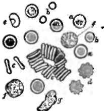
Fig. 60. Human Blood Corpuscles, a, a. Red corpuscles of the ordinary appearance; b, b, red corpuscles of smaller size, such as are now and then seen exceptionally; c, c, red corpuscles seen in profile, single and in rows; d, two red corpuscles shrivelled by partial evaporation of the serum; e, a red corpuscle swollen by slight addition of water; f,f, white corpuscles; g, one which has undergone amoeboid change of form.
These observations furnish a rough but instructive analysis of blood. Three constituents are exhibited, the corpuscles, the serum, and the fibrin. The serum and fibrin together constitute the liquor sanguinis, or blood plasma. The blood may thus be separated into liquor sanguinis and corpuscles, or into defibrin-ated blood and fibrin, or, lastly, into serum and a coloured clot consisting of the fibrin with the corpuscles entangled in it.

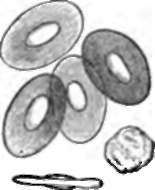
Fig. 61. Blood Corpuscles of Frog, viz., four red corpuscles seen in full view, one in profile, and one white corpuscle.
Continue to:
